Chapter: 11 th 12th std standard Bio Botany plant tree Biology Higher secondary school College Notes
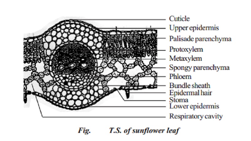
Anatomy of a dicot leaf - Sunflower leaf
Anatomy of a dicot and monocot leaves
Leaves are very important vegetative organs because they are mainly concerned with photosynthesis and transpiration. Like stem and roots, leaves also have the three tissue systems - dermal, ground and vascular. The dermal tissue system consists of an upper epidermis and lower epidermis. Stomata occur in both the epidermis but more frequently in the lower epidermis. The ground tissue system that lies between the epidermal layers of leaf is known as mesophyll tissue. Often it is differentiated into palisade parenchyma on the adaxial (upper) side and spongy parenchyma on the abaxial (lower) side.
A leaf showing this differentiation in mesophyll is designated as dorsiventral. It is common in dicot leaves. If mesophyll is not differentiated like this in a leaf (i.e., made of only spongy or palisade parenchyma) as in monocots, it is called isobilateral. The mesophyll tissue, especially spongy parenchyma cells enclose a lot of air spaces. The presence of air spaces is a special feature of spongy cells. They facilitate the gaseous exchange between the internal photosynthetic tissue (mesophyll) and the external atmosphere through the stomata.
The vascular tissue system is composed of vascular bundles. They are collateral and closed. The vascular tissue forms the skeleton of the leaf and they are known as veins. The veins supply water and minerals to the photosynthetic tissue. Thus the morphological and anatomical features of the leaf help in its physiological functions.
Anatomy of a dicot leaf - Sunflower leaf
Internal structure of dicotyledonous leaves reveals epidermis, mesophyll and vascular tissues.
Epidermis
A dicotyledonous leaf is generally dorsiventral. It has upper and lower epidermis. The epidermis is usually made up of a single layer of cells that are closely packed. The cuticle on the upper epidermis is thicker than that of lower epidermis. The minute openings found on the epidermis are called stomata. Stomata are more in number on the lower epidermis than on the upper epidermis. A stoma is surrounded by a pair of bean shaped cells called guard cells.
Each stoma opens into an air chamber. These guard cells contain chloroplasts, whereas other epidermal cells do not contain chloroplasts. The main function of the epidermis is to give protection to the inner tissue called mesophyll. The cuticle helps to check transpiration. Stomata are used for transpiration and gas exchange.
Mesophyll
The entire tissue between the upper and lower epidermis is called the mesophyll (Gk meso=in the middle; phyllome=leaf). There are two regions in the mesophyll. They are palisade parenchyma and spongy parenchyma. Palisade parenchyma cells are seen beneath the upper epidermis. It consists of vertically elongated cylindrical cells in one or more layers. These cells are compactly arranged without intercellular spaces. Palisade parenchyma cells contain more chloroplasts than the spongy parenchyma cells. The function of palisade parenchyma is photosynthesis. Spongy parenchyma lies below the palisade parenchyma. Spongy cells are irregularly shaped. These cells are very loosely arranged with numerous airspaces. As compared to palisade cells, the spongy cells contain lesser number of chloroplasts. Spongy cells facilitate the exchange of gases with the help of air spaces. The air space that is found next to the stoma is called respiratory cavity or sub-stomatal cavity.
Vascular tissues
Vascular tissues are present in the veins of leaf. Vascular bundles are conjoint, collateral and closed. Xylem is present towards the upper epidermis, while the phloem towards the lower epidermis. Vascular bundles are surrounded by a compact layer of parenchymatous cells called bundle sheath or border parenchyma. Xylem consists of metaxylem vessels and protoxylem vessels. Protoxylem vessels are present towards the upper epidermis. Phloem consists of sieve tubes, companion cells and phloem parenchyma. Phloem fibres are absent. Xylem consists of vessels and xylem parenchyma. Tracheids and xylem fibres are absent.
Related Topics