Chapter: Human Nervous System and Sensory Organs : Diencephalon
Specific Thalamic Nuclei - Dorsal Thalamus

Specific Thalamic Nuclei
The cortex-dependentspecific thalamic nu-clei are subdivided into the following
nuclear groups (or complexes):
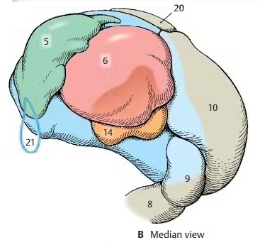
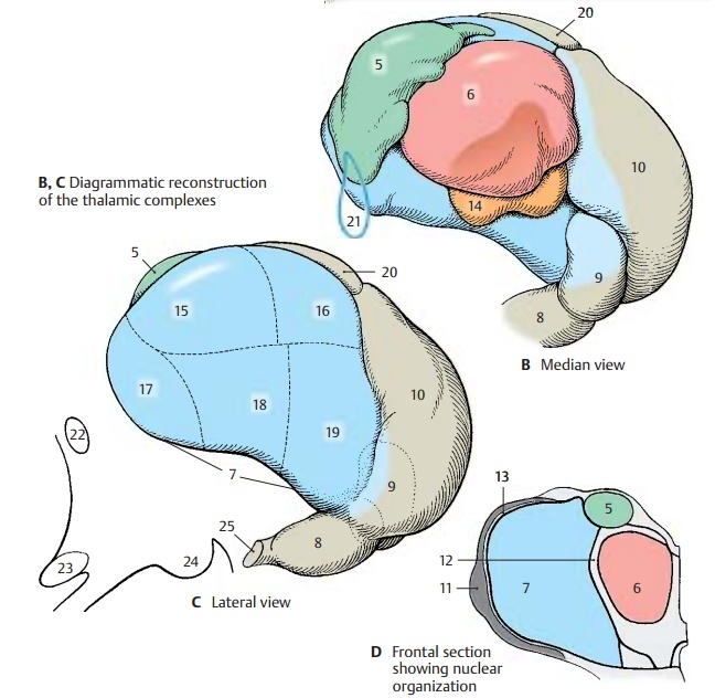
! The anterior nuclear group, anteriorthalamic nuclei (green) (B –
D5).
! The
medial nuclear group, medial
thalamicnuclei (red) (BD6).
!
The lateral nuclear group, ventrolateralthalamic nuclei (blue) (CD7); this group isfurther divided into a lateral tier, the lateral nuclei, and a ventral tier, the ven-tral nuclei
! The
lateral geniculate nucleus (BC8).
! The medial geniculate nucleus (BC9).
! The pulvinar (BC10).
!
The reticular
nucleus of the thalamus (D11).
The
nuclear groups are separated by layers of fibers: the internal medullary lamina (D12)
(between the medial nuclear group and the lateral and anterior nuclear groups)
and the external medullary lamina (D13) (between thelateral nuclear group
and the reticular nu-cleus which encloses the lateral surface of the thalamus).
The
reticular nucleus and nonspecific nu-clei, with the exception of the centromediannucleus (B14), have been omitted from
thereconstruction of nuclear groups (B,
C). The most anterior nuclear groups
are the ante-rior nuclei (B5) to
which the medial nuclei (B6) border
caudally. In the lateral complex, we distinguish between a nuclear group
lo-cated dorsally (the lateral nuclei, lateral
dor-sal nucleus [C15] and lateral posterior nu-cleus [C16]), and a group located
ventrally(the ventral nuclei, ventral
anterior nucleus [C17], ventral lateral nucleus [C18], and ven-tral posterior nucleus [C19])
BC20
Superficial dorsal nucleus.
B21
Interventricular foramen (foramen of Monro).
C22
Anterior commissure.
C23
Optic chiasm.
C24
Mamillary body.
C25
Optic tract.

Each nuclear group is connected to a specific region (field of projection) in the cerebral cortex; hence the term specificthalamic nuclei. Within this system, thenuclei project to their cortical fields and, in turn, the cortical fields project to the re-spective thalamic nuclei. Thus, there exists a neuronal circuit with a thalamocortical limb and a corticothalamic limb. The neu-rons of the specific thalamic nuclei transmit impulses to the cerebral cortex and are, in turn, influenced by the respective cortical fields. Hence, the function of a cortical field cannot be examined without the thalamic nucleus belonging to it; likewise, the func-tion of a thalamic nucleus cannot be ex-amined without the cortical field belonging to it.
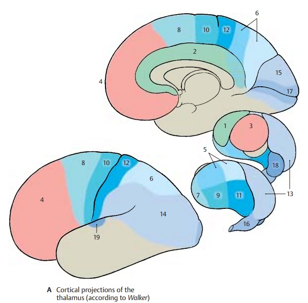
When neurons of the specific thalamic nu-clei become separated from their axon ter-minals, they respond by retrograde degeneration. Therefore, destruction of cir-cumscribed cortical fields results in neu-ronal death in the respective thalamic nu-clei. The thalamic projection fields of the cerebral cortex can be delineated in this way. The anterior nuclei (A1) are connected with the cortex of the cingulate gyrus (A2) and the medial nuclei (A3) with the cortex of the frontal lobe (A4). The lateral nuclei (A5) project to the dorsal and medial cortex of the parietal lobe (A6), with the lateral dorsal nucleus partly supplying the retrosplenial part of the cingulate gyrus. Among the ven-tral nuclei, the ventral anterior nucleus (A7) is connected with the premotor cortex (A8), the ventral lateral nucleus (A9) with the motorprecentral area (A10), and theventral poste-rior nucleus (A11) with thesensory postcen-tral area (A12). Thepulvinar(A13) projects tothe cortical parts of the parietal and tem-poral lobes (A14) and to the cuneus (A15).The lateral geniculate nucleus (A16) is con-nected through the visual pathway with the visual cortex (striate area) (A17), themedialgeniculate nucleus (A18) through the auditorypathway with the auditory cortex (trans-verse temporal gyri, Heschl’s convolutions) (A19)..

Related Topics