Chapter: Human Nervous System and Sensory Organs : Diencephalon
Lateral Geniculate Body - Dorsal Thalamus
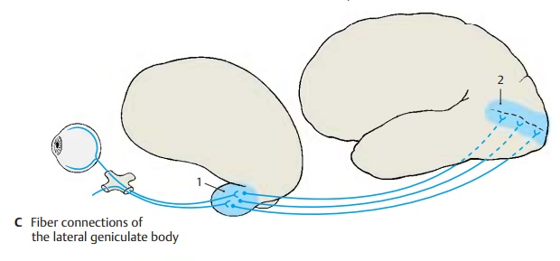
Lateral Geniculate Body
This
nucleus (C1) lies somewhat isolated
at the ventrocaudal aspect of the thalamus and is a relatively independent
structure. It shows stratification into six cell layers which are separated by
the afferent fiber bundles of the optic
tract. Crossed and un-crossed optic fibers terminate in a regular arrangement
in each of the two geniculate nuclei. In the left lateral genicu-late body, the
temporal half of the retina of the left eye and the nasal half of the retina of
the right eye are represented; in the right lateral geniculate body, the
temporal half of the retina of the right eye and the nasal half of the retina
of the left eye are represented. The fibers from the macula, which is the region of greatest visual acuity, termi-nate
in a central wedge-shaped area, which extends through all cell layers. The neurons
of the lateral geniculate nu-cleus send their axons to the visual cortex, the striate area (C2) at the medial hemispheric surface of the occipital lobe (central optic radiation or occipitothalamicradiation).

Related Topics