Chapter: Human Nervous System and Sensory Organs : Diencephalon
Frontal Section Through the Tuber Cinereum
Frontal Section Through the Tuber Cinereum
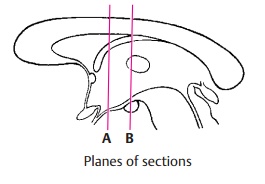
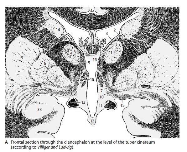
The plane of section lies just behind the interventricular foramen (foramen of Monro). Lateral ventricle and third ventricle are separated by the thin base of the choroid plexus (A1). From here to the thalamostriate vein (AB2) extends the lamina affixa (AB3). It covers the dorsal surface of the thalamus (A4), of which only the anterior nuclei are visible. Ventrolaterally to it and separated by the internal capsule (AB5) lies the globus pallidus (AB6), which is divided into two parts, the internal (A7) and the external seg- ment of the pallidum (A8). It stands out against the adjacent putamen (AB9) because of its higher myelin content. At the basal margin and at the tip of the pallidum there exit the lenticular fasciculus (Forel’s fieldH2) and the lenticular ansa (A10). The latter forms an arch in dorsal direction around the medial tip of the pallidum. The ventral part of the diencephalon is occupied by the hy- pothalamus (tuber cinereum [A11] and in- fundibulum [A12]), which appears markedly poor in myelin, in contrast to the heavily myelinated optic tract (AB13). The diencephalon is enclosed on both sides by the telencephalon, but without a clearly visible boundary. The closest nuclei of the telencephalon are the putamen (AB9) and the caudate nucleus (AB14). Anterior to theglobus pallidus lies a nucleus belonging to the telencephalon, the basal nucleus (Mey-nert’s nucleus) (A15). It receives fibers fromthe midbrain tegmentum. Its large choliner-gic neurons project diffusely into the entire neocortex. The fornix (AB16) is seen twice because of its arched course.


Related Topics