Chapter: Human Nervous System and Sensory Organs : Diencephalon
Frontal Section Through the Rostral Thalamus
Frontal Section Through the Rostral Thalamus
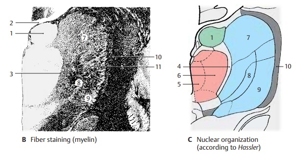
In the myelin staining, the
anterior and me-dial nuclear groups are clearly distinguishable from the
lateral nuclear group by their poor and delicate myelination. The dorsally
located anterior nuclear group
(green) (A – C1) bulges against the interventricular foramen (foramen of Monro)
(AB2) and forms the thalamic eminence. Themedial nuclear group (red) is enveloped
by the internal medullarylamina (B3) and the intralaminar nuclei (C4)which separate it from the lateral
portion. Within the medial nuclear group, a medial magnocellular portion (AC5) and a lateral parvocellular
portion (AC6) surrounding the medial
one can be distinguished.
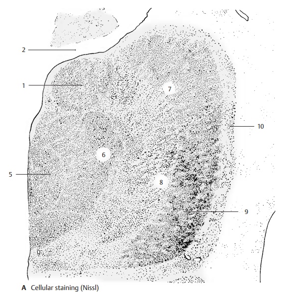
The largest part of the thalamus
is formed by the lateroventral nuclear
group (blue), which surrounds the medial portion like a broad shell. It
contains considerably more myelin, and a difference between its dorsal and
ventral regions can be recognized in the myelin stained section (B). As compared to the dorsal region
(lateral dorsal nucleus) (A – C7), the ventral nuclear region has
more prominent and coarser myelin fibers. Its di-vision into a medial and a
lateral segment can be easily recognized in the overview. The section shows the
ventral lateral nu-cleus. In its medial segment (A–C8) termi-nate fibers
from the midbrain tegmentum. Lateral to it is seen the rostral part of the
nu-cleus (A – C9) where the fiber bundles of the superior cerebellar peduncle
terminate; its projection to the precentral area (area 4) re-veals a
somatotopic organization.
The
lateral surface of the thalamus is formed by the reticular nucleus of the thalamus (A – C10). As a narrow
layer of cells, this nucleus laterally surrounds the entire thalamus like a
shell and extends from the rostral pole, where it is widest, to the pulvi-nar
and to the lateral geniculate nucleus. It is separated from the lateral nuclear
com-plex by a lamella of myelin fibers, the exter-nal
medullary lamina (B11). The
relation-ships between cerebral cortex and reticular nucleus vary for different
nuclear segments: the frontal cortex is connected with the ros-tral portion of
the nucleus, the temporal cortex with the middle portion, and the occipital
cortex with the caudal portion. The functional significance of this nucleus is
un-known. Its neurons send many collaterals to the other thalamic nuclei.

Fiber
relationships between the thalamic nuclei and certain cortical areas have been
established by experimentally destroying the cortical segments or by severing
the fibers. The neurons of the respective nuclei undergo retrograde degeneration
once their axons have been severed. Neurons of the re-ticular nucleus, however,
are thought to un-dergo transneuronal degeneration rather than retrograde
degeneration, that is, they not degenerate because their axons have been
severed but because they have lost af-ferent fibers terminating on them. This
would mean that the cortex projects to the reticular nucleus, while the latter
does not project to the cortex.
Related Topics