Chapter: Human Nervous System and Sensory Organs : Diencephalon
Frontal Section Through the Caudal Thalamus
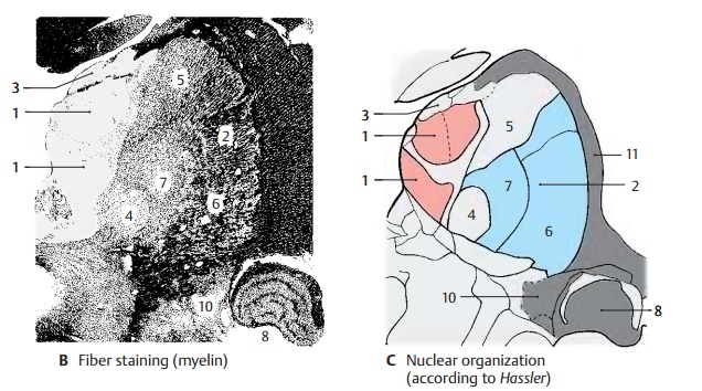
Frontal Section Through the Caudal Thalamus
At this
level, the section again shows the medial
nuclear group (red) (A – C1) and the lateroventral nuclear group (blue) (A – C2). Itincludes the
most caudal parts of the medial nucleus. Dorsally separated from the super-ficial dorsal nucleus (A–C3)
by a narrow my-elin layer, they are otherwise surrounded by the internal
medullary lamina and the in-tralaminar nuclei. The nonspecific thalamic parts
here reach special expansion through the centromedian
nucleus (A – C4).
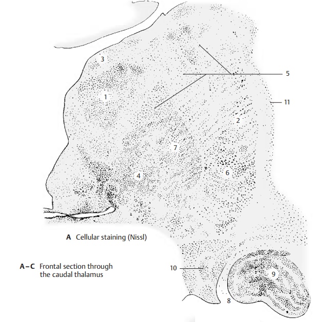
The most
rostral nuclear portions of the pulvinar
(A – C5) lie dorsally between themedial and the lateral nuclear groups.
This rostral part of the pulvinar projects to the upper convolutions of the
temporal lobe and is thought to receive fibers from the lateral lemniscus; it is therefore assumed tobe an integration
nucleus of the acoustic system.
The ventral posterior nucleus (A – C6) is seen in the lateroventral area. The medial lemnis-cus, the spinothalamic pathways, and thesecondary
trigeminal fibers terminate here. The outer portion, which receives the fibers
for the limbs and the trunk, is rich in myeli-nated fibers and has fewer cells
than the inner portion, which receives the fibers for the head region. The
inner portion is rich in cells and has thinly myelinated fibers. It sur-rounds
the centromedian nucleus ventrally and laterally; it appears as a
crescent-shaped figure in the myelin-stained section and, hence, is called the semilunar nucleus (B7).

The lateral geniculate body (A – C8) lies slightly apart from the complex of the thalamus at the
ventral surface of the dien-cephalon. It is indented at the base and pro-trudes
laterally (lateral geniculum). It is
characterized by prominent stratification into six layers of cells and five
intercalating layers of fibers. The latter are formed by the fibers of the optic tract, which disperse ac-cording
to a set pattern and terminate at neurons of different cell layers. The upper
four of these layers are parvo-cellular, the lower two layers are
magno-cellular. In the second, third, and fifth layers terminate the fibers
from the retina of the ipsilateral eye (uncrossed optic nerve fibers), while
those from the contralateral eye (crossed optic nerve fibers) terminate in the
first, fourth, and sixth layers. Fibers from the site of visual acuity, the
macula, termi-nate in the central area (A9).
When the mac-ula is destroyed, the geniculate cells of this area undergo
transneuronal degeneration. The lateral geniculate body is surrounded by a
dense capsule of myelinated fibers. These are the dorsally and laterally
emerging fibers of the optic radiation
(geniculocal-carine tract).
Medially
to the lateral geniculate body, the section shows the caudal portion of the me-dial geniculate body (A–C10).
The reticular nucleus (AC11) forms the lateral capsule.
Itwidens ventrally and also encloses the lateral geniculate body.
Related Topics