Chapter: Essentials of Anatomy and Physiology: Respiratory System
Pleural Cavities - Anatomy of the Respiratory System
Pleural Cavities
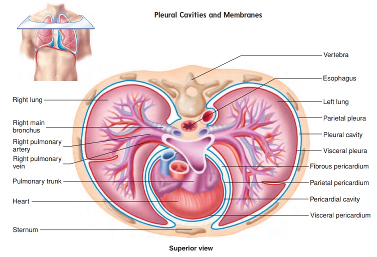
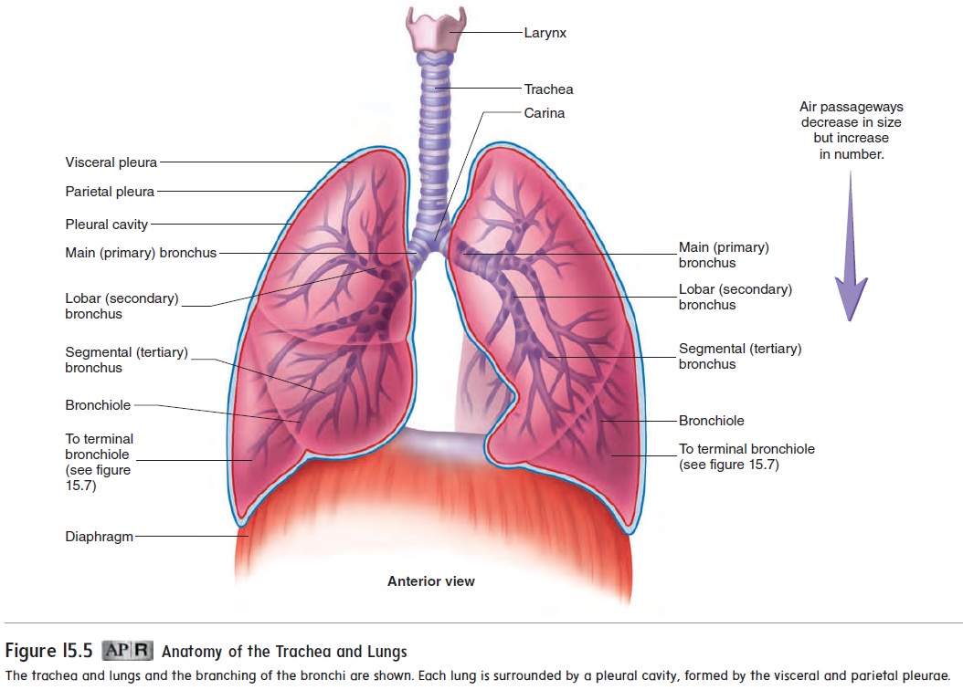
The lungs are contained within the thoracic cavity. In addition, each lung is surrounded by a separate pleural (ploor′ ăl; relating to the ribs) cavity. Each pleural cavity is lined with a serous membrane called the pleura. The pleura consists of a parietal and a visceral part. The parietal pleura, which lines the walls of the thorax, dia-phragm, and mediastinum, is continuous with the visceral pleura, which covers the surface of the lung (figure 15.9; see figure 15.5).
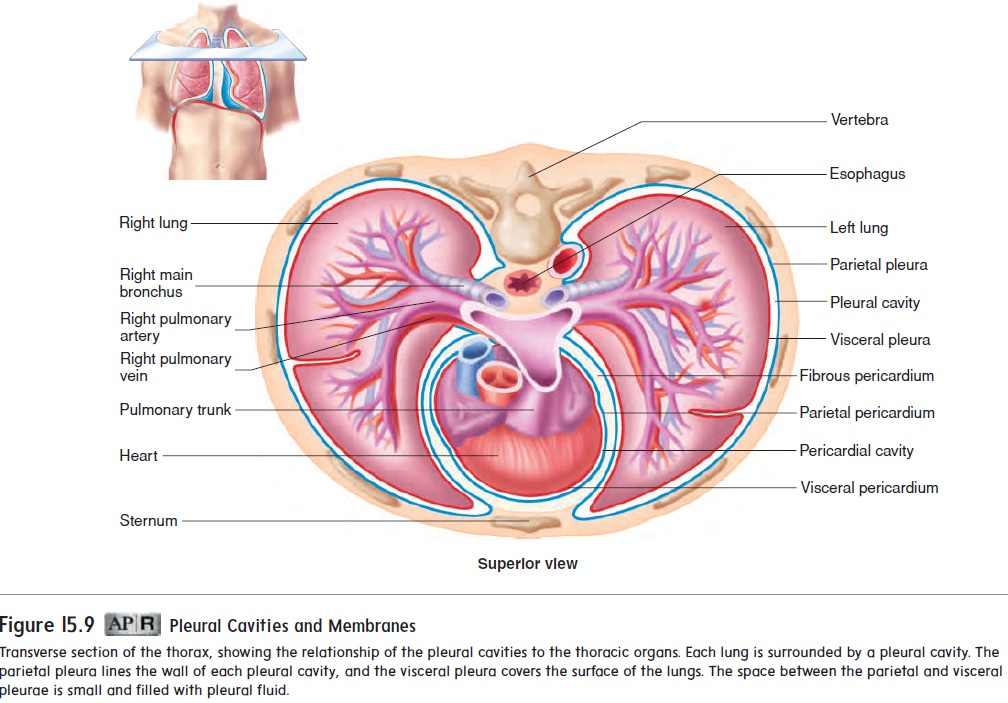
The pleural cavity, between the parietal and visceral pleurae, is filled with a small volume of pleural fluid produced by the pleural membranes. The pleural fluid performs two functions:
(1) It acts as a lubricant, allowing the visceral and parietal pleurae to slide past each other as the lungs and thorax change shape during respiration, and (2) it helps hold the pleural membranes together. The pleural fluid acts similarly to a thin film of water between two sheets of glass (the visceral and parietal pleurae); the glass sheets can slide over each other easily, but it is difficult to separate them.
Related Topics