Chapter: Essentials of Anatomy and Physiology: Respiratory System
Larynx - Anatomy of the Respiratory System
Larynx
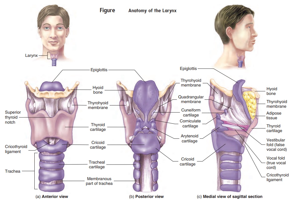
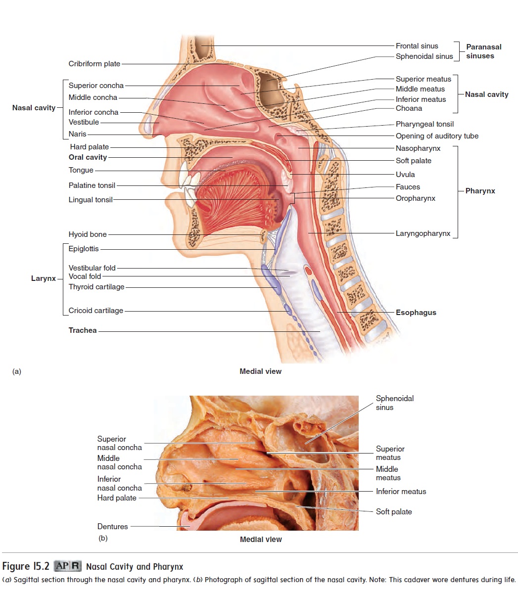
The larynx (lar′ ingks) is located in the anterior throat and extends from the base of the tongue to the trachea (figure 15.2a). It is a pas-sageway for air between the pharynx and the trachea. The larynx consists of an outer casing of nine cartilages connected to one another by muscles and ligaments (figure 15.3). Three of the nine cartilages are unpaired, and six of them form three pairs. The largest cartilage is the unpaired thyroid (thı̄′ royd; shield-shaped) cartilage, or Adam’s apple. The thyroid cartilage is attached superiorly to the hyoid bone. The most inferior cartilage of the larynx is the unpaired cricoid (krı̄′koyd; ring-shaped) cartilage, which forms the base ofthe larynx on which the other cartilages rest. The thyroid and cricoid cartilages maintain an open passageway for air movement.
The third unpaired cartilage is the epiglottis (ep-i-glot′ is; on the glottis). It differs from the other cartilages in that it consists of elastic cartilage rather than hyaline cartilage. Its inferior margin is attached to the thyroid cartilage anteriorly, and the superior part of the epiglottis projects superiorly as a free flap toward the tongue. The epiglottis helps prevent swallowed materials from entering the larynx. As the larynx elevates during swallowing, the epiglottis tips posteriorly to cover the opening of the larynx.
The six paired cartilages consist of three cartilages on each side of the posterior part of the larynx (figure 15.3b). The top carti-lage on each side is the cuneiform (kū′ nē-i-fōrm; wedge-shaped) cartilage, the middle cartilage is the corniculate (kōr-nik′ū-lāt;horn-shaped) cartilage, and the bottom cartilage is the arytenoid (ar-i-tē′ noyd; ladle-shaped) cartilage. The arytenoid cartilages articulate with the cricoid cartilage inferiorly. The paired carti-lages form an attachment site for the vocal folds.
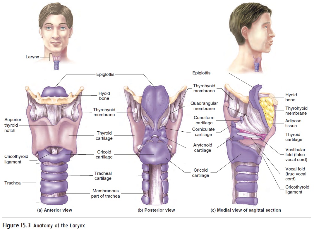
Two pairs of ligaments extend from the posterior surface of the thyroid cartilage to the paired cartilages. The superior pair forms the vestibular (ves-tib′ ū-lăr) folds, or false vocal cords, and the inferior pair composes the vocal folds, or true vocal cords (figure 15.4). When the vestibular folds come together, they pre-vent air from leaving the lungs, as when a person holds his or her breath. Along with the epiglottis, the vestibular folds also prevent food and liquids from entering the larynx.
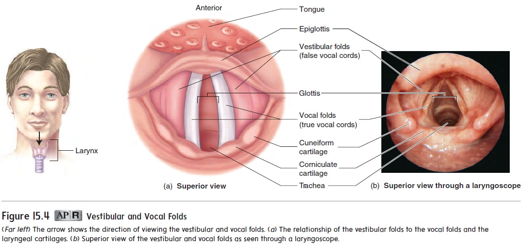
The vocal folds are the primary source of voice production. Air moving past the vocal folds causes them to vibrate, producing sound. Muscles control the length and tension of the vocal folds. The force of air moving past the vocal folds controls the loudness, and the tension of the vocal folds controls the pitch of the voice. An inflammation of the mucous epithelium of the vocal folds is called laryngitis (lar-in-jı̄′ tis). Swelling of the vocal folds during laryngitis inhibits voice production.
Related Topics