Chapter: Ophthalmology: Optic Nerve
Optic Nerve
Optic Nerve
Basic Knowledge
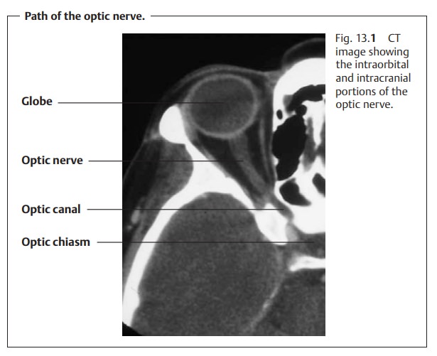
The optic nerve extends from the posterior
pole of the eye to the optic chiasm
(Fig. 13.1). After this
characteristic crossing, the fibers of the optic nerve travel as the optic tract to the lateral geniculate body. Depending on the shape of the skull, the
optic nerve has a total length of 35 – 55 mm. The nerve consists of:
An intraocular portion.
An
intraorbital portion.
An
intracranial portion.
Intraocular Portion of the Optic Nerve
The intraocular portion of the optic nerve is
visible on ophthalmoscopy as the optic disk. All the retinal nerve fibers merge into the optic nerve here,
and thecentral retinal vessels enter and leave the eye here. The complete
absence of photoreceptors at this site creates a gap in the visual field known
as the blindspot.
Shape and size:
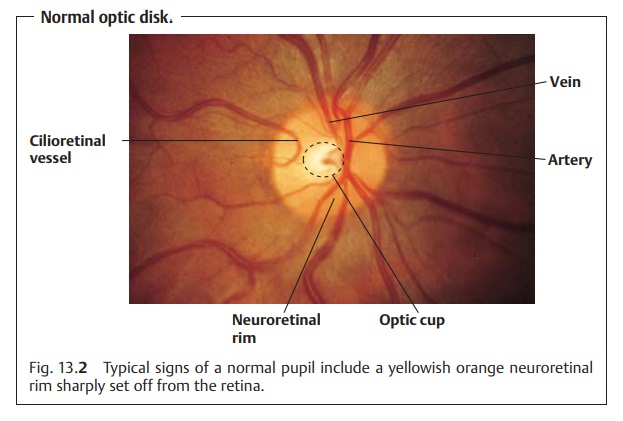
The optic disk (Fig. 13.2)
is normallyslightly vertically ovalwith
an average area of approximately 2.7 mm2 and a horizontal diameter of approximately 1.8 mm. There is a wide range of physiologic variability in
thesize of the optic disk; its area may vary by a factor of seven, and its
horizontaldiameter by a factor of two and one-half.
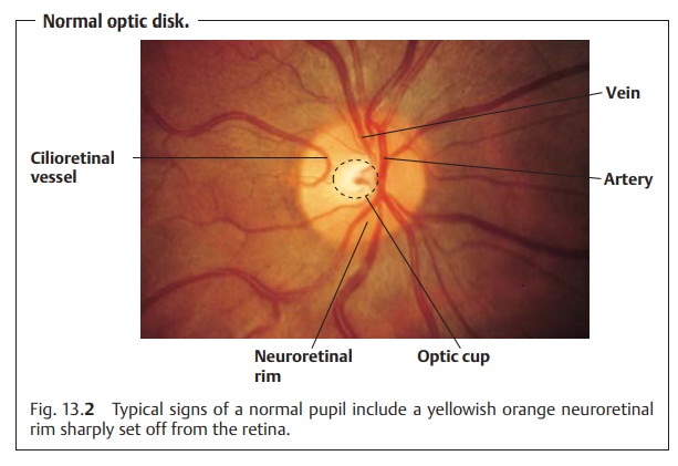
Color:
The normal physiologic color isyellowish orange. The temporal half ofthe optic disk is usually
slightly paler.
Margin:
The margin of the optic disk issharply definedand readily distin-guished from the surrounding
retinal tissue. On the nasal side, the greater density of the nerve fibers
makes the margin slightly less distinct than on the temporal side. A common
clinical observation is a crescent of pigment or irregular pigmentation close
to the optic disk on the temporal side; some-times the sclera will be visible
through this crescent.
Prominence of the optic disk:
The normal optic disk is not prominent. Thenerve fibers are practically flush with the retina.
Neuroretinal rim (Fig. 13.2):
This
consists of the bundles of all the optic nervefibers as they exit through the
scleral canal. The rim has a characteristic
con-figuration: The narrowest portion is in the temporal horizontal region
fol-lowed by the nasal horizontal area; the widest areas are the vertical
inferior and superior areas.
Optic cup:
This is theslightly
eccentric cavitationof the optic nerve that has aslightly flattened oval
shape corresponding to that of the neuroretinal rim. It is the brightest part
of the optic disk. No nerve fibers exit from it (Fig. 13.2). The size of the optic
cup correlates with the size of the optic disk; the larger the optic disk,
the larger the optic cup. Because enlargement of the optic cup means a loss of
nerve fibers in the rim, it is particularly
important to documentthe size of the optic cup. This is specified as the
horizontal and vertical ratios of cup to
disk diameter (cup/disk ratio). Due to the wide range of variability
inoptic disk size, it is not possible to specify absolute cup/disk ratios that
indi-cate the presence of abnormal processes.
Central retinal artery and vein:
These structures usually enter the eyeslightly nasal to the
center of the optic disk. Visible pulsation in the vein is normal. However, arterial pulsation is always abnormal and occurs with
dis-orders such as increased intraocular pressure and aortic stenosis.
Cilioretinal vessels are aberrant vessels originating directly from the choroid(short
posterior ciliary arteries). Resembling a cane, they usually course along the
temporal margin of the optic disk and supply the inner layers of the retina
(Fig. 13.2).
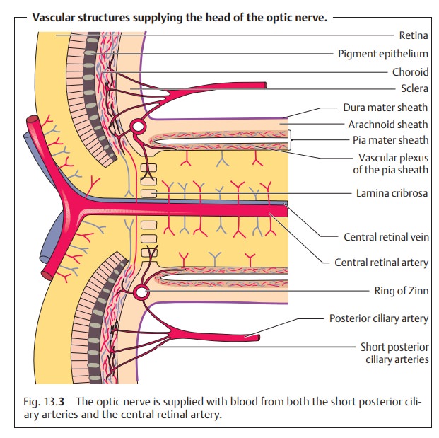
Blood supply to the optic disk (Fig. 13.3):
The
optic disk receives its bloodsupply from the ring of Zinn, an anastomotic ring
of small branches of the short posterior ciliary arteries and the central
retinal artery. Both groups of vessels originate from the ophthalmic artery,
which branches off of the inter-nal carotid artery and enters the eye through
the optic canal. The central reti-nal artery and vein branch into the optic
nerve approximately 8 mm before the point at which the optic nerve exits the
globe. Approximately 10 short posterior ciliary arteries penetrate the sclera
around the optic nerve.
The Intraorbital and Intracranial Portion of the Optic Nerve
The intraorbital portion begins after the nerve passes through a
sieve-like plateofscleralconnectivetissue,thelaminacribrosa.
Insidetheorbit,theoptic nerve describes an S-shaped course that allows extreme
eye movements.
After the optic nerve passes through the optic
canal, the short intracranialportion begins and extends as far as the optic chiasm.
Like the brain, theintraorbital and intracranial portions of the optic nerve
are surrounded by sheaths of dura mater, pia, and arachnoid (see Fig. 13.3). The nerve receives its blood supply
through the vascular pia sheath.
Related Topics