Chapter: Ophthalmology: Optic Nerve
Intraocular Optic Nerve Tumors

Tumors
Optic nerve tumors are classified as intraocular or retrobulbar tumors.
Intraocular tumors are rare.
Intraocular Optic Nerve Tumors
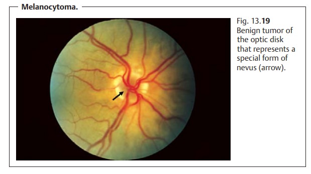
Melanocytoma (Fig. 13.19): These are benign pigmented tumors that pri-marily occur in
blacks. The color of the tumor varies from gray to pitch black. It is often
eccentric and extends beyond the margin of the optic disk. In 50% of all cases,
one will also observe a peripapillary choroidal nevus. Visual acuity is usually
normal, although discrete changes in the visual field my be present.

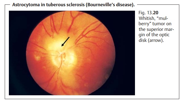
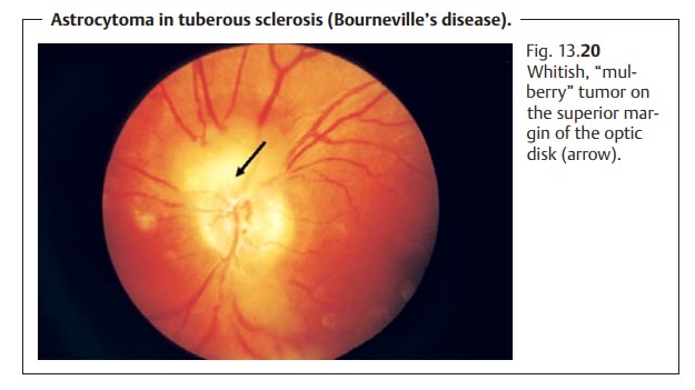
Astrocytoma (Fig. 13.20): Astrocytomas appear as white reflecting “mul-berry” masses that can calcify. Their size can range up to several disk diame-ters. The tumor is highly vascularized. Visual field defects can result where the tumor is sufficiently large to compress the optic nerve. Astrocytomas occur in tuberous sclerosis (Bourneville’s disease) and neurofibromatosis (Recklinghausen’s disease).

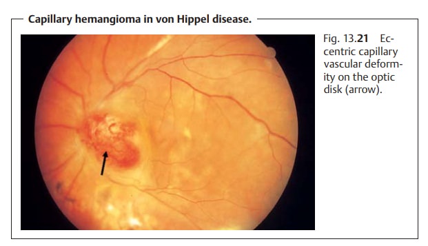
Hemangioma (Fig. 13.21): Capillary hemangiomas are eccentric, roundorange-colored
vascular deformities on the optic disk (von Hippel disease). They may occur in
association with other angiomas, for example in the cere-bellum (in von
Hippel-Lindau disease).

Related Topics