Chapter: Ophthalmology: Optic Nerve
Arteriosclerotic Anterior Ischemic Optic Neuropathy
Arteriosclerotic Anterior Ischemic Optic Neuropathy
Definition
An acute disruption of the blood supply to the
optic disk, i.e., optic disk infarc-tion, resulting from vascular changes in
arteriosclerosis.
Epidemiology:
Arteriosclerotic AION is a common cause of
sudden loss of visual acuity. The greatest incidence of this disorder is between
the ages of 60 and 70. In contrast to arteritic AION, it can also occur in
adults below the age of 60.
Etiology:
The causes of the disorder lie in acute disruption of the blood flowthrough the lateral branches of the short posterior ciliary arteries and the ring of Zinn in the setting of severe arteriosclerosis. A narrow scleral canal, i.e., a small optic disk, is a predisposing factor. The disorder known as diabeticpapillopathy also belongs to this group of disorders, although it has a betterprognosis in terms of vision.
Symptoms:
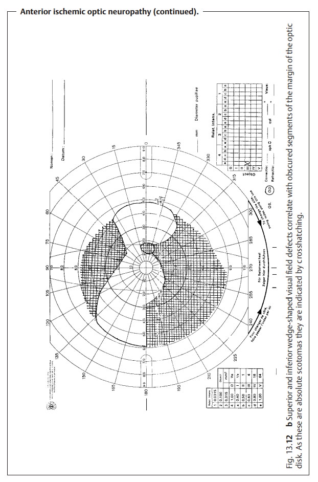
Patients report asudden unilateral loss of visual acuity. This is dueto segmental or
complete infarction of the anterior portion of the optic nerve. Severity is
variable. The patient may present with wedge-shaped visual field defects (Fig.
13.12b) or horizontal visual field
defects that correlate with seg-mental nerve fiber edemas. However, severe
concentric defects progressing to total blindness can also occur. Vision may or
not be impaired. An afferent pupillary defect is always present.
Diagnostic considerations:
The patient will frequently have a history
ofhypertension, diabetes mellitus, or hyperlipidemia.
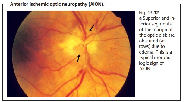
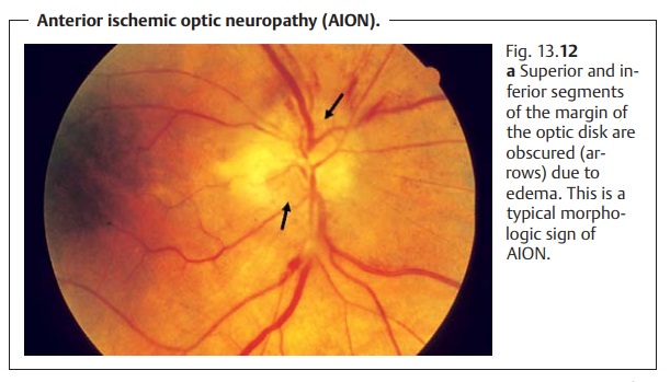
Ophthalmoscopy will reveal edema of the optic disk, whose margin willbe
accordingly obscured. The margin is often obscured in a segmental pat-tern,
which is an important criterion in differential diagnosis (Fig. 13.12a). The head of the optic nerve is
also hyperemic with marginal bleeding.
Obscured segments of the margin of the optic
disk that correlate with visual field defects are a sign of AION.
Treatment:
Anterior ischemic optic neuropathy is nearly impossible to treat.Attempted methods include hemodilution (pentoxifylline infusions, acetyl-salicylic acid, and bloodletting depending on hematocrit levels) and systemic administration of steroids to control the edema. Diagnosis of the underlying cause is important; examination by an internist and Doppler ultrasound stud-ies of the carotid artery may be helpful. Underlying disorders such as diabetes mellitus or arterial hypertension should be treated.
Prognosis:
The prognosis is usually poor even where
therapy is initiatedearly. Isolated atrophy of the optic nerve will appear
within three weeks, complex atrophy of the optic nerve is less frequent but may
also be observed.
Related Topics