Chapter: Human Nervous System and Sensory Organs : Telencephalon
Neocortex: Fiber Tracts

Fiber Tracts
A broad layer of white matter lies between the cerebral cortex and the gray nuclei in the depth. It consists of fiber bundles originating from the neurons of the cortex or those extending to the cortex and terminating on the cortical neurons. There are three different types of fiber systems:
! Projection fibers
! Association fibers
! Commissural fibers
Projection fibers provide connections be-tween the cerebral cortex and the subcorti-cal centers, either as ascending systems ter-minating in the cortex or as descending sys-tems extending from the cortex to the deeper-lying centers. Association fibers pro-vide connections between different cortical areas of the same hemisphere. Commissuralfibers connect the cortices of both hemi-spheres; they are really nothing else but in-terhemispheric association fibers. However, commissural fibers often connect identical regions of the two hemispheres.
Projection Fibers
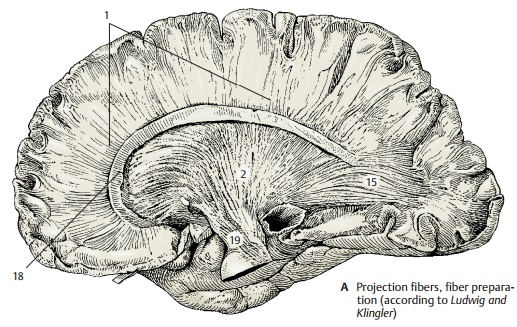
The pathways descending from different cortical areas merge and form a fanlike structure known as the internal capsule. The ascending fibers pass through the inter-nal capsule and then radiate outward like a fan. In this way, ascending and descending fibers form a radiating crown of fibers beneath the cortex, the corona radiata (A1).
The internal capsule (A2, B) appears in hor-izontal sections as an angle consisting of an anterior limb (B3), bordered by the head ofthe caudate nucleus (B4), globus pallidus (B5) and putamen (B6), and a posterior limb (B7), bordered by the thalamus (B8), globus pallidus and putamen (see p. 222, A). Be-tween both limbs lies the genu of the internalcapsule (B9). The different fiber tracts passthrough specific parts of the internal cap-sule. The frontopontine tract (B10) (red lines) and the anterior thalamic radiation (B11) (blue lines) pass through the anterior limb. The corticonuclear fibers supplying thecranial nerve nuclei run through the genu of the internal capsule. The fibers of the corti-cospinal tract (red dots) pass through theposterior limb in a somatotopic organiza-tion: upper extremity, trunk, and lower ex-tremity. The thalamocortical fibers leading to area 4 and the corticorubral and corti-cotegmental fibers coming from area 6 pass through the same region. The caudal part of the posterior limb is occupied by the fibers of the central thalamic radiation (blue dots) (B12) which extend to the postcentral area. The fibers of the posterior thalamic radiation(B13) (light blue dots) and of the tem-poropontine tract (light red dots) (B14) passobliquely through the caudal part.
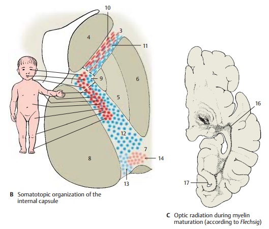
The most important projection pathways include the acoustic radiation and the optic radiation. The fibers of the acoustic radia-tion originate in the medial geniculatebody, extend across the lateral geniculate body, and cross the internal capsule at the inferior margin of the putamen. In the white matter of the temporal lobe, they ascend al-most vertically to the anterior transverse gyrus (Heschl’s convolution) (pp. 252, 380). The optic radiationoriginates in the lateral geniculate body. The fibers fan out into a wide medullary lamina (A15) and run to the temporal lobe, where they form the tem-poral genu (C16) of the visual pathway. They then pass in an arch around the inferior horn of the lateral ventricle and through the white matter of the occipital lobe to the cal-carine sulcus (C17).
A18 Corpus callosum.
A19 Cerebral peduncle.
Association Fibers (A – D)
The connections between different cortical areas are very different in length. For the purpose of simplification, we distinguish short and long association fibers.
The short association fibers, or arcuatefibers (B), provide connections within acerebral lobe (B1) or from one convolution to the next (B2). The shortest fibers connect adjacent cortical parts. They reenter the cortex after running just a short distance through the white matter. The layer of ar-cuate fibers lies immediately beneath the cortex.
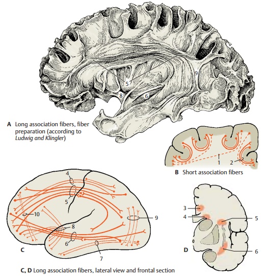
The long association fibers connect differ-ent cerebral lobes and form compact bundles that can be recognized with the naked eye. The cingulum(D3) is a strong sys-tem of shorter and longer fibers lying un-derneath the cingulate gyrus; it follows the entire course of the cingulate gyrus. The long fibers extend from the parolfactory area and the rostrum of the corpus callosum to the entorhinal area. The subcallosalfasciculus (superior occipitofrontal fasciculus) (CD4) lies dorsolaterally to thecaudate nucleus below the radiation of the corpus callosum. Its fibers connect the fron-tal lobe with the temporal lobe and the occipital lobe. Some of the fibers run to the insula, others connect the frontal lobe with the caudate nucleus. Thesuperior longitudi-nal fasciculus (ACD5) lying dorsolaterally tothe putamen is a strong association bundle between frontal lobe and occipital lobe with fibers branching to the parietal lobe and temporal lobe. The inferior occipitofrontalfasciculus (ACD6) passes through the ventralpart of the extreme capsule from the frontal lobe to the occipital lobe. The inferior longi-tudinal fasciculus (C7) extends betweenoccipital lobe and temporal lobe. The unci-nate fasciculus (AC8) connects the temporalcortex with the frontal cortex. Its ventral part provides a connection between the en-torhinal area and the orbital area of the frontal lobe. Other fiber bundles are the ver-tical occipital fasciculus (AC9) and the orbi-tofrontal fasciculus (C10).
Commissural Fibers (E, F)
The interhemispheric association fibers pass through the corpus callosum, the anterior commissure, and the commissure of the fornix to the con-tralateral hemisphere. The most important commissure of the neocortex is the corpuscallosum (E). Its curved rostral part is thegenu of the corpus callosum (E11) with thepointed rostrum (E12). It is followed by the middle part, the trunk of the corpus callosum (E13), and the thickened end, thespleniumof the corpus callosum (E14). The fibers ofthe corpus callosum spread through the white matter of both hemispheres and form the radiation of the corpus callosum. The arched fibers passing through the genu of the corpus callosum and connecting both frontal lobes are called minor forceps(F15), those passing through the splenium and connecting both occipital lobes are called major forceps (F16).
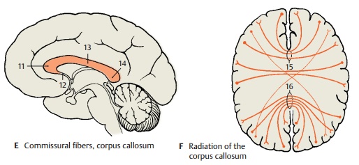
We distinguish between homotopic and heterotopic interhemispheric fibers. Homo-topic fibers connect the same cortical areasin both hemispheres, while heterotopic fibersconnect different areas. The vast majority of fibers of the corpus callosum is homotopic. Not all areas are connected to the same ex-tent with their counterpart in the other hemisphere. The hand and foot parts of both somatosensory areas, for example, possess no interhemispheric fiber connections; nor are the two visual cortices connected to each other. However, very strong fiber con-nections exist between both areas 18, which are regarded as optic integration areas.
Related Topics