Chapter: Human Nervous System and Sensory Organs : Telencephalon
Insula

Insula
The insula is the region at the lateral
aspect of the hemisphere that lags behind during development and becomes
covered by the more rapidly growing adjacent regions of the hemisphere. The
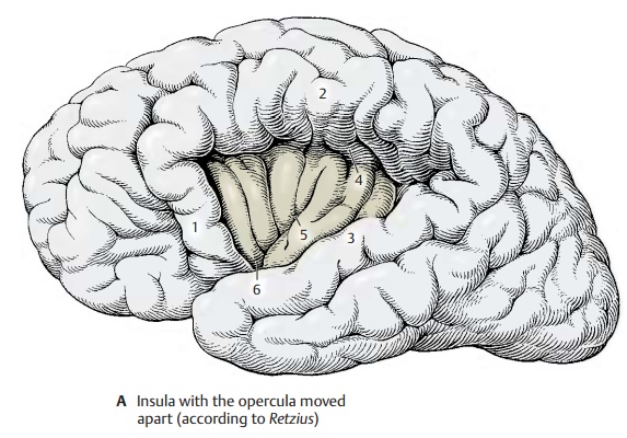
parts of the hemisphere overlapping the insula are called opercula. They are named according to thecerebral lobe they belong
to: the frontaloperculum (A1), the parietal operculum (A2),and
the temporal operculum (A3). In diagram A, the opercula have been moved apart toexpose the insula. They
normally leave only a cleft, the lateral
cerebral sulcus (fissure ofSylvius,
p. 10, A4), which widens over the in-sula into the lateral fossa. The insula has roughly the shape of a triangle and
is bordered at its three sides by the circular
sulcus of the insula (A4). Thecentralsulcus of the insula (A5) divides the insulainto a rostral
and a caudal part. At its lower pole, the limen
of insula (A6), the insular
re-gion merges into the olfactory area, the paleocortex.

The
insular cortex represents a transitionalregion
between paleocortex and neocortex.The lower pole of the insula is occupied
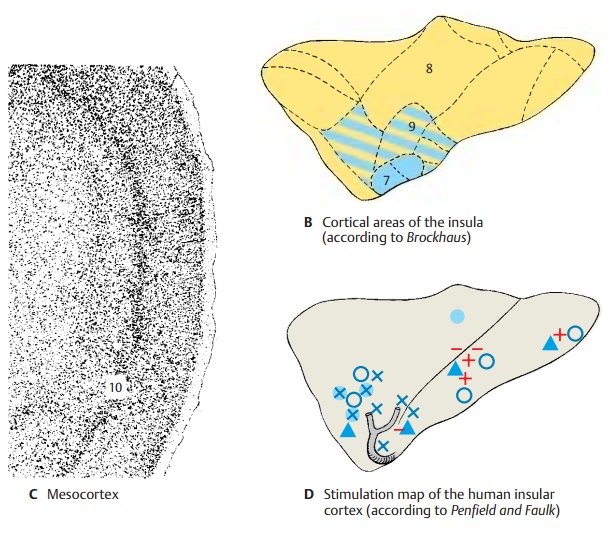
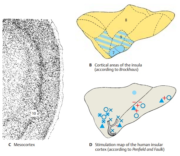
by the prepiriform area (B7) (blue) which belongs to the
paleocortex. The upper part of the insula is covered by the isocortex (neo-cortex; see p. 244) (B8) (yellow) with the fa-miliar six
layers. Between both parts lies a transitional region, the mesocor-tex (proisocortex, see p. 244) (B9) (hatchedarea). Unlike the paleocortex, it has six lay-ers;
however, these are only poorly developed as compared to the neocortex. The
fifth layer (C10) is characteristic
for the mesocortex by standing out as a distinct narrow, dark stripe in the
cortical band. It contains small pyramidal cells that are densely packed like
palisades, a feature otherwise found only in the cortex of the cingulate gyrus.

Stimulation responses (D).Stimulation
ofthe insular cortex is difficult because of the hidden position of the region;
it has been carried out in humans during surgical treat-ment of some specific
forms of epilepsy. Itcaused an increase (+) or decrease ( ŌĆō ) in the
peristaltic movement of the stomach. Nausea and vomiting (├Ė) were induced at
some stimulation sites, while sensations in the upper abdomen or stomach region
(x) or in the lower abdomen (o) were produced at other
sites. At several stimulation sites, taste sensations were induced (Ōłå).
Al-though the stimulation chart does not show a topical organization of these
effects, the results do indicate viscerosensory
andvisceromotor functions of the insular cortex.Experiments with monkeys
yielded not only salivation but also motor responses in the muscles of the face
and the limbs. In humans, surgical removal of the insular re-gion does not lead
to any functional losses.
Related Topics