Chapter: Human Nervous System and Sensory Organs : Telencephalon
Neostriatum

Neostriatum
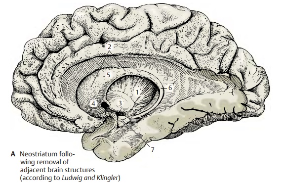
The neostriatum (or striatum) is the highestintegration site of the extrapyramidal motor system. It is a large, gray complex inthe depth of the cerebral hemisphere and is divided into two parts by the internal cap-sule (ABD1), namely, the caudate nucleus (ABD2) and the putamen (ABD3). The caudate nucleus consists of the large head ofthe caudate nucleus (A4), the body of the cau-date nucleus (A5), and the tail of the caudate nucleus(A6). Immunohistochemical assaysfor neurotransmitter substances yield a spotty, mosaic-like structure created by the terminals of various fiber tracts. The spots form a system of interconnected fields (striosomes) that stand out from the rest of the tissue because of their content of a specific neurotransmitter.

Afferent Pathways (B ‚Äď D)
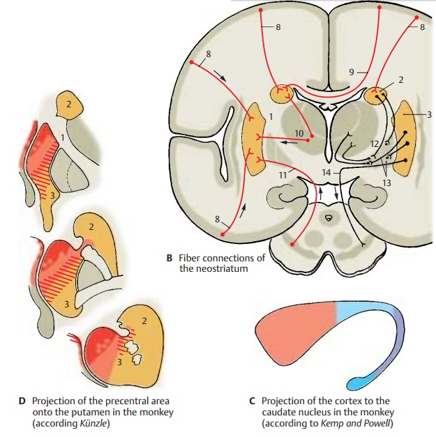
Corticostriate fibers (B8). Fibers extendfrom all areas of the neocortex to the neo-striatum. They are the axons of medium-sized and small pyramidal cells of the fifth layer (see p. 240). However, there are no fiber connections extending from the stri-atum to the cortex. The corticostriate pro-jection reveals a topical organization (C): the frontal lobe projects to the head of the caudate nucleus (red) and is followed by the parietal lobe (light blue), the occipital lobe (purple), and the temporal lobe (dark blue) (see p. 213). The projection of the precentral motor area in the putamen reveals a soma-totopic organization (D): head (red), arm (light red), and leg (hatched area). A soma-totopic projection of the postcentral sensory area to the dorsolateral region of the caudate nucleus has been demon-strated. The fibers from areas adjoining the central sulcus are the only ones that partly cross via the corpus callosum to the con-tralateral neostriatum (B9).
Centrostriate fibers (B10). These fiberbundles extend from the centromedian thalamic nucleus to the neostriatum; those for the caudate nucleus originate in the dor-sal part, those for the putamen in the ven-tral part of the nucleus. Impulses from the cerebellum and from the reticular forma-tion of the midbrain reach the neostriatum via these fibers.
Nigrostriate fibers (B11). Fibers extendingfrom the substantia nigra to the neostri-atum can be traced by fluorescence micros-copy. They are the axons of dopaminergic neurons, and they cross the inner capsule in groups. They run without interruption through the globus pallidus to the neostri-atum.
Serotoninergic fiber bundles from theraphe nuclei.
Efferent Pathways (B)
The efferent fibers extend to the globus pal-lidus. The fibers of the caudate nucleus ter-minate in the dorsal parts of the two seg-ments of the pallidum (B12), while the fibers of the putamen terminate in the ven-tral parts (B13). Here, they synapse with the pallidofugal system, namely, with the pal-lidosubthalamic fibers, the lenticular ansa, the lenticular fasciculus, and the pal-lidotegmental fibers.
Strionigral fibers (B14). Fibers of the cau-date nucleus terminate in the rostral part and fibers of the putamen in the caudal part of the substantia nigra.

Functional Significance
Both the topical organization of the cor-ticostriate fiber systems and its mosaic-like structure show that the neostriatum is divided into many functionally different sectors. It receives stimuli from the frontal cortex, from the optic, acoustic, and tactile cortical fields and their association areas. These areas are thought to have an effect on the motor system via the stratum (sensory motor integration, cognitive function of the neostriatum). The neostriatum has no direct control over elementary motor processes (its destruction does not lead to an appreci-able loss of motor functions). Rather, it is viewed as a higher integration system that influences the behavior of an individual.
A7 Amygdaloid body.
Related Topics