Chapter: Human Nervous System and Sensory Organs : Telencephalon
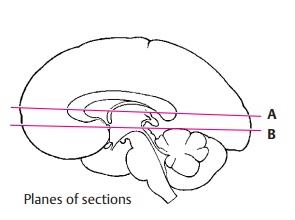
Horizontal Sections of Brain Through the Telencephalon
Horizontal Sections
1. Superior Aspect of Corpus Callosum and Lateral Ventricles,
2. Exposure of the Roof of the Diencephalon,
3. Horizontal Section through the Neostriatum ,
4. Exposure of the Roof of the Diencephalon.
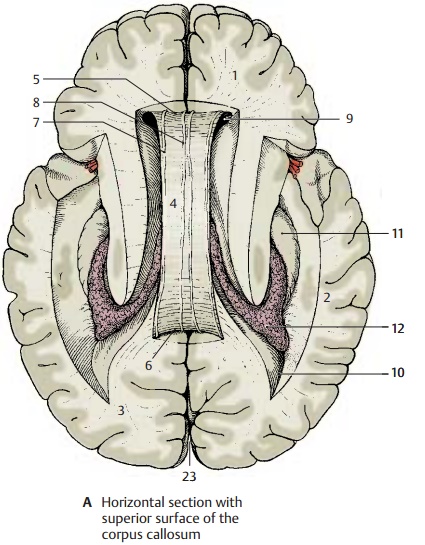
Superior Aspect of Corpus Callosum and Lateral Ventricles (A)
The horizontal section through the brain has been cut above the corpus callosum, and the superior aspect of the corpus callosum and the lateral ventricles have been exposed by removal of deeper portions of white mat-ter. The section shows the frontal lobes (A1) at the top, the temporal lobes (A2) on both sides, and the occipital lobes (A3) at the bottom. The superior surface of the corpus callosum (A4) belongs to the free brain sur-face lined by the pia mater and arachnoidea. Lying deep in the brain, it is covered by the convolutions of the medial walls of the hemispheres. Rostrally, the superior surface of the corpus callosum turns in ventral direction and forms the genu of the corpuscallosum (A5); caudally, itforms the splenium of the corpus callosum (A6). On the superior aspect of the corpus callosum extend four myelinated fiber ridges: one lateral longitudinal stria (A7) and one medial longitudinal stria ofLancisi (A8) run along each half of the cor-pus callosum (see p. 230). Their fiber tracts extend from the hippocampus to the sub-callosal area. Between the two longitudinal striae lies a thin layer of gray matter con-sisting of a narrow layer of neurons, the in-dusium griseum. This is a cortical portion ofthe archicortex that regressed as a result of the extensive development of the corpus callosum and subsequent displace-ment of the archicortex into the inferior horn of the lateral ventricle (see p. 209, F).
The anterior horns (A9) of the lateral ven-tricles are opened in the area of the frontal lobes, and the posterior horns (A10) in the area of the occipital lobes. The protruding hippocampus (A11) forms the floor of the inferior horn. The central part and the inferior horn of the lateral ventricle contain the choroid plexus (A12).
Exposure of the Roof of the Diencephalon (B)
This is an oblique horizontal section below the corpus callosum, which has been completely removed. Upon opening the two lateral ventricles, the dorsal aspect of the caudate nucleus (B13) and, bordering medi-ally, the dorsal aspect of the thalamus (B14) become visible. Parts of the diencephalon become exposed as well, namely, the pinealgland (B15) and both habenulae (B16) whichare connected to it. The two fornices (B17) between the heads of the two caudate nu-clei have been cut in their rostral part (columns of fornix). The septum pellucidum (B18) extends from there to the corpus callosum.
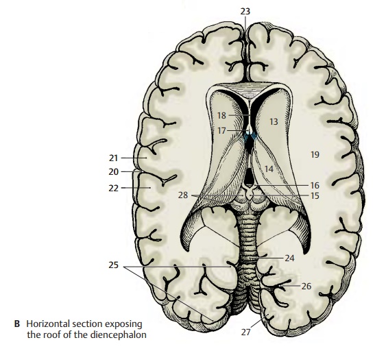
The lateral wall of the hemisphere contains a particularly wide medullary layer be-tween the cortex and the ventricle, the semioval center (B19). The central sulcus (B20) cuts into it and separates the frontal lobe (at the top of the figure) from the parietal lobe (bottom). Starting from the central sulcus, the precentral gyrus (B21) and the postcentral gyrus (B22) can be lo-cated. Caudally in thelongitudinal cerebral fissure
(AB23), the cerebellum (B24) is visible. The caudal portion of the hemisphere is formed by the occipital lobe. The striate area (B25), the visual cortex, lies in this region and oc-cupies primarily the calcarine sulcus (B26) at the medial aspect of the occipital lobe, while extending only a short distance onto the occipital pole. It can be distinguished even by the naked eye from the rest of the cortex through a white streak, the line ofGennari (B27), which divides the cortex into two gray bands. Gennari’s line is a wide band of myelinated nerve fibers corresponding to the slightly narrower external band of Baillarger in the other areas of the neocortex .
Horizontal Section through the Neostriatum (A)
At this level, the lateral cerebral fossa (AB1) is exposed in its longitudinal expansion. The lateral sulcus (A2) is found more rostrally,with the frontal operculum (AB3) in front of it and the elongated temporal operculum (AB4) caudally to it. The longitudinal expan-sion is also apparent in the deep structures of the telencephalon, the claustrum (AB5) and the putamen (AB6). The arched struc-tures have been cut twice; the corpus callo-sum (A7) appears rostrally with its anteriorpart, the genu of the corpus callosum, and caudally with its end, the splenium. The cau-date nucleus has been cut twice as well; the head of the caudate nucleus (AB8) is seenrostrally and the tail of the caudate nucleus (AB9) caudolaterally to the thalamus (AB10). The thalamus is separated from theglobus pallidus (AB11) by the internal cap-sule which, in horizontal sections, exhibitsthe shape of a hook made up of theanteriorlimb (AB12) and the posterior limb (AB13).Also the lateral ventricle has been exposed twice. Its anterior horn (A14) has been cut in the area of the frontal lobe and, caudally, in the transition to the posterior horn (A15). The two anterior horns are separated by the septum pellucidum (A16), which spans be-tween corpus callosum and fornix (A17).
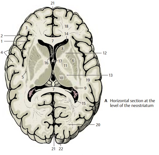
The section also shows the frontal lobes (AB18), the temporal lobes (AB19), the occipital lobes (A20), the longitudinal cere-bral fissure (AB21), and the striate area (visual cortex) (A22).
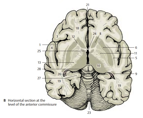
Horizontal Section at the Level of the Anterior Commissure (B)
While the section still shows the entire frontal lobe and temporal lobe, the occipital lobe has only been cut in its anterior part at the transition to the temporal lobe. Between the two hemispheres appears the cone-shaped dorsal aspect of the cerebellum (B23). The anterior horn of the lateral ven-tricle and the corpus callosum are no longer seen in this section. Instead there is theanterior commissure (B24) connecting thetwo hemispheres. The two columns of thefornix (B25), lying close together in the pre-vious section, are separated at the level of the anterior commissure. While the poste-rior limb of the internal capsule (AB13) re-tains its usual width, the anterior limb (AB12) is only indicated by some fiber bundles. As a result, the head of the caudatenucleus (AB8) is no longer separated fromthe putamen (AB6), and the striatum is seen as uniform nuclear complex. In the area of the temporal lobe, the curled-up cortical band of the hippocampus (Ammon’s horn) (B26) is almost covered by the parahippo-campal gyrus (B27).

Related Topics