Chapter: Human Nervous System and Sensory Organs : Telencephalon
Archicortex's Subdivision and Functional Significance

Subdivision and Functional Significance
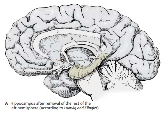
The hippocampus (A – D1) is the main part of the archicortex. It lies at the medial aspect of the temporal lobe in the depth and is largely covered by the parahippocampal gyrus. The left hemisphere has been removed in the preparation, showing the cut surface of the corpus callosum (A2) with only the left hippocampus being left intact. The latter looks like a paw with claws, the digitations. The temporal lobe of the righthemisphere in the background illustrates the position of the hippocampus in the tem-poral lobe. The hippocampus extends to the caudal end of the corpus callosum. Here, it becomes reduced to a thin layer of gray matter, the indusium griseum (A3), which ex-tends along the superior surface of the cor-pus callosum to its rostral end in the region of the anterior commissure (A4). Two nar-row fiber bundles, the lateral and the mediallongitudinal striae of Lancisi also run here bilaterally. On the dorsal surface of the hippocampus lies a thick fiber band, the fimbria of hippocampus (A – D5), which separates from the hippocampus beneath the corpus callosum and continues as fornix (A6), arching down to to mamillarybody (A7).
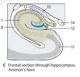
In a horizontal section through the temporal lobe, the inferior horn (BC8) and the poste-rior horn (B9) of the lateral ventricle are ex-posed, and the protrusion of the hippocam-pus into the ventricle is visible. Medially, al-ready at the outer aspect of the temporal lobe, lies the fimbria and, beneath it, the dentate gyrus (fascia dentata) (B – D10), sepa-rated from the parahippocampal gyrus (en-torhinal area) (B – D11) by the hippocampalsulcus (BC12).
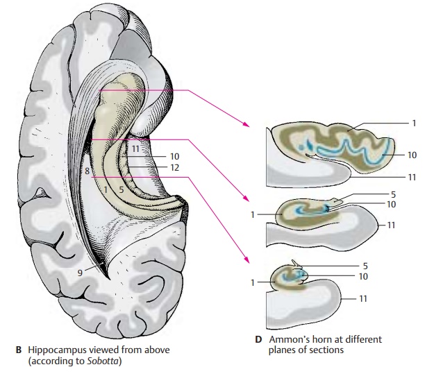
In a frontal section the hippocampal cortex forms a curled band, Ammon’s horn, which protrudes against the ventricle and is covered by a layer of fibers, the alveus (C13). Ammon’s horn shows considerable varia-tions at different planes of section (D)In the past, the hippocampus has been as-signed to the rhinencephalon, but it has no direct relationship with the olfactory sense. In reptiles, which do not have a neocortex, the telencephalon is the highest integration organ. Electrical recordings from the hippo-campus of mammals show that it receives optic, acoustic, tactile, and visceral input, but only a few olfactory impulses. It is an integration organ influencing endocrine, visceral, and emotional processes via its con-nections to the hypothalamus, septal nuclei, and cingulate gyrus. Furthermore, the hip-pocampus plays a major role in processes of learning and memory.
Clinical Note: Bilateral removal of the hippo-campus in humans (treatment of severe epileptic seizures) leads to a loss in memory. While old memories are retained, new information can be remembered only for a few seconds. Such a short-term memory may persist for years. The hippo-campal neurons possess a very low absolute threshold for convulsive discharges. Thus, the hippocampus is of special importance for the origin of epileptic seizures and memory deficits.
C14 Optic tract.
C15 Choroid plexus.
Related Topics