Chapter: Human Nervous System and Sensory Organs : Telencephalon
Neocortex: Frontal Lobe

Frontal Lobe
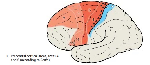
We distinguish the precentral area (the motor cortex proper) and the premotor, the prefrontal, and the orbitofrontal cortical areas.
Agranular Cortex
The cortex of the precentral area (red), con-sisting of primary motor cortex (area 4) and premotor cortex (area 6) (C), is characterizedby the reduction or loss of granular layers and a general increase in pyramidal cells. Also typical are the exceptional thickness of the cortex and its gradual transition into the white matter. These features are especially prominent in the cortex of area 4 (A), where certain regions of layer V contain giant pyra-midal cells (Betz’s cells) (A1). The latterpossess the thickest and longest axons in the nervous system, reaching as far as the sacral spinal cord.
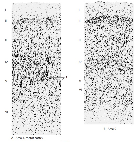
The prefrontal cortex (area 9, light red) is shown for comparison (B). It is not only nar-rower and better delimited against the white matter by a distinct layer VI, but also possesses well-developed granular layers (II and IV).
The agranular cortex (areas 4 and 6) is the principal site of origin of the pyramidal tract and is regarded as prototype of the motorcortex. Nevertheless, it also receives afferentfibers: following the stimulation of skin at the extensor and flexor sides of the limbs, electrical potentials can be recorded at the precentral area. They are probably afferent systems for controlling and fine-tuning the motor system. On the other hand, motor re-sponses can be induced by enhanced stimu-lation at some points of the postcentral area of the parietal lobe (somatosensory area, blue) (C) and the premotor area of the fron-tal lobe. Accordingly, pyramidal cells with long axons are found in layer V of these areas (!). Physiologists therefore speak of a motosensory area, Ms I (predominantlymotor) and of a sensorimotor area, Sm I (pre-dominantly sensory). However, these find-ings do not affect the basic fact that the pre-central area represents the motor cortex,while the postcentral area represents the somatosensory (tactosensory) cortex.
Granular Cortex
The prefrontal and orbitofrontal cortices exhibit well-developed granular layers (B).
Clinical Note: Injury to the granular frontalcortex results in severe changes in personality. This affects the formal intellectual capacity less than it does initiative, ambition, concentration, and judgment. The patients show a silly self-satisfied euphoria, are only interested in everyday trivia, and are unable to plan ahead.
Similar changes have been observed in patients with prefrontal lobotomy. This is a complete surgi-cal severing of the frontal fiber connections, which was performed as a form of treatment of raving, mentally deranged patients and in patients with the most severe conditions of pain (this treatment has now become obsolete thanks to psychopharmaceuticals). The operation achieved permanent calming and indifference of the patients. A characteristic change was ob-served in the emotional sphere: the patents still felt the pain, but they did not perceive it as trou-blesome; previously intolerable pain had become immaterial. Radical changes in character also occur following injury of the orbitofrontal area (basal neocortex). Previously cultured and edu-cated people exhibit a disintegration of decency, tact, and sense of shame, which may lead to seri-ous social offenses.
Somatotopic Organization of the Precentral Area (A, B)
Electrical stimulation of individual cortical regions of the precentral area (A, B1) causes muscle contraction in specific body regions. The area exhibits a somatotopic organiza-tion. The head region lies above the lateral sulcus, with the lowest parts representing the throat (A2), the tongue (A3), and the lips. The areas for hand, arm, trunk, and leg follow dorsally, with the leg area reaching beyond the edge of the pallium to the me-dian surface of the hemisphere. This creates an inverted motor homunculus. The areas for individual body parts differ in size; those parts of the body where muscles have to perform differentiated movements are rep-resented by particularly large areas. The fin-gers and the hand occupy the largest areas, and the trunk the smallest.
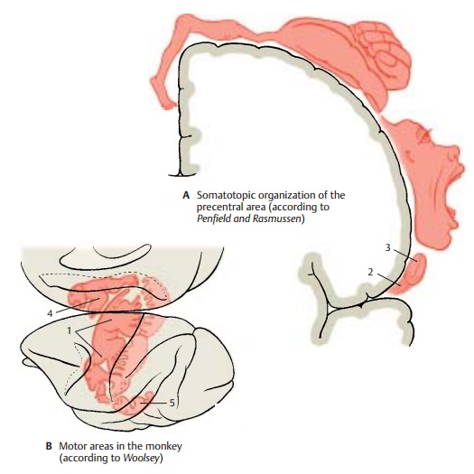
Each body half is represented on the con-tralateral hemisphere, that is, the left body half on the right hemisphere and the right body half on the left hemisphere. Unilateral stimulation yields bilateral responses of masticatory, laryngeal, and palatal muscles, and partly also of the trunk muscles. The muscles of face and limbs yield strictly con-tralateral responses. Representation of thelimbs is organized in such a way that areas for distal parts of the limbs lie deep in the central sulcus, while those for the proximal parts lie more rostrally on the precentral gyrus (B1).
Supplementary Motor Areas (B)
Apart from the precentral area (Ms I), there are two additional motor areas. Their soma-totopic organization has been demon-strated in monkeys but not in humans. The second motosensory area, Ms II (B4), lies onthe medial surface of the hemisphere above the cingulate gyrus adjacent to areas 4 and 6. The second sensorimotor area, Sm II (B5), which is predominantly a tactosensory area rather than a motor area, lies above the lateral sulcus and roughly corresponds to area 40. The functional significance of theseareas within the entire motor system has not yet been elucidated.
Frontal Eye Field (C)
Conjugated eye movements can be induced by electrical stimulation of the precentral area and, above all, of area 8. This is the fron-tal eye field for voluntary eye movements. In general, stimulation causes the gaze to turn to the opposite side, sometimes with simultaneous turning of the head. The fibers of area 8 do not terminate directly at the nuclei of the eye-muscle nerves. Their im-pulses are probably relayed in the intersti-tial nucleus of Cajal.

Broca’s Motor Speech Area (D)
Injury to the region in the lower frontal gyrus (areas 44 and 45) of the dominant hemisphere causes motor aphasia. The patients are no longer able to form and articulate words, even though the speech muscles (lips, tongue, larynx) are not paralyzed. Speech comprehension is not af-fected.
It is certainly impossible to localize the faculty of speech to a defined cortical area (“speech center”). Speech is one of the highest cerebral functions and involves large regions of the cortex. However, Broca’s motor speech area is without doubt a crucial relay station in the complex neuronal foundation of speech
Related Topics