Chapter: Basic Radiology : Radiology of the Chest
Exercise: Multiple Pulmonary Nodules
EXERCISE 4-8.
MULTIPLE PULMONARY NODULES
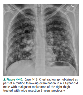
4-13. The most likely cause of the multiple pulmonary nodules
in Case 4-13 (Figure 4-40) is metastasis. herpes simplex pneumonia.
Radiologic Findings
4-13. In this case, the chest radiograph shows multiple,
smoothly marginated, solid nodules in both lungs. These nodules are distributed
diffusely and have vari-ous diameters (A is the correct answer to Question
4-13). The heart is normal in size and shape.
Discussion
The radiographic pattern of
multiple pulmonary nodules is frequently encountered (Table 4-8). The clinical
setting has considerable influence on the differential diagnosis in such cases
and should always be taken into account when assess-ing patients with this
pattern. However, the differential diag-nosis may be narrowed by assessing the
absolute size of the nodules, the uniformity of their size, their marginal
charac-teristics, whether or not they are calcified, and whether or not they
are cavitary. In adults the most common causes of multiple nodules are
metastatic neoplasm and infectious disease. Metastatic neoplasm may result from
carcinoma, sarcoma, or lymphoma. Pulmonary metastases may be of any size and
number. In contrast to inflammatory nodules, nodular pulmonary metastases are
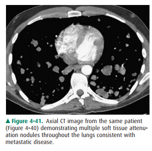
often of various diame-ters. Metastases are usually of soft-tissue density
similar to muscle or blood (Figure 4-41). Metastases may rarely be cal-cified
if the patient has a sarcoma that makes bone or carti-lage (eg, osteosarcoma).
Differentiation is most commonly made by the clinical setting or review of old
studies, but de-termination of the correct diagnosis may require tissue biopsy
for confirmation.

Multiple pulmonary nodules may
also be due to infec-tious disease, most commonly fungal or mycobacterial
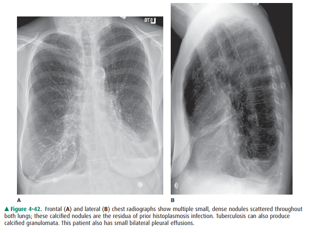
infec-tions. In the United States, the most common fungus is histoplasmosis
(Figure 4-42), although there are regional variations. Calcified nodules that
are all of similar size sug-gest a previous infection with either
histoplasmosis or tuber-culosis. Nodules seen in acute infection are often not
as sharply defined as metastases. This is especially true if the nodules
represent acinar shadows. In these instances, the nodule is approximately 5 to
10 mm in diameter and is ill de-fined or fuzzy on its margin. Acinar nodules
develop in pa-tients with viral pneumonias such as herpes pneumonia or chicken
pox (varicella) pneumonia.
Multiple pulmonary nodules may
also develop in a wide variety of other disorders, including Wegener’s
granulomato-sis and arteriovenous malformations, but would not be as numerous
as in this case.
Related Topics