Chapter: 11 th 12th std standard Bio Botany plant tree Biology Higher secondary school College Notes
Development of male and female gametophyte

Development of male and female gametophyte
The stamen or microsporophyll consists of a filament, anther and a connective. Each anther consists of two anther lobes connected by a midrib known as connective. Each anther lobe contains two pollen sacs or microsporangia.
Development of microsporangium
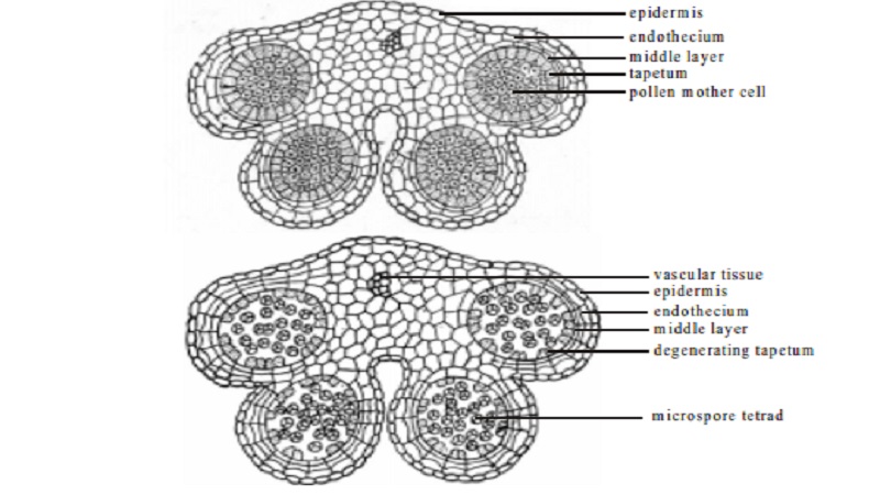
The cross section of a very young anther shows a mass of undifferentiated cells surrounded by epidermis. The rows of hypodermal cells called microsporangial initial or archesporium becomes differentiated in each lobe of the anther.
Each microsporangial initial divides by periclinal division to form outer primary parietal cell and inner primary sporogenous cell. The primary parietal cell repeatedly divides to form the wall layers as described below :
A. Epidermis
It is the outermost layer of a young anther and undergoes only anticlinal divisions.
B. Endothecium
Immediately below the epidermis, the cell layers are radially elongated and develop fibrous thickenings. These cells are hygroscopic in nature and help in dehiscence.
C. Middle layer
Usually one to three middle layers are found below the endothecium. They become crushed at the time of meiotic division in the pollen.
D. Tapetum
The cells of the innermost parietal layer possess dense protoplasm and the food entering into the sporogenous cells pass through it. Thus it serves as a nutritive layer for the developing microspore. The tapetum may be glandular or amoeboid based on the behaviour of the cells during sporogenesis.
E. Sporogenous tissue
The primary sporogenous cells undergo several divisions to form microspore mother cells. Each microspore mother cell divides meiotically to produce four microspores or pollengrains which will have half the (n) number of chromosomes.
F. Microspore or pollengrain
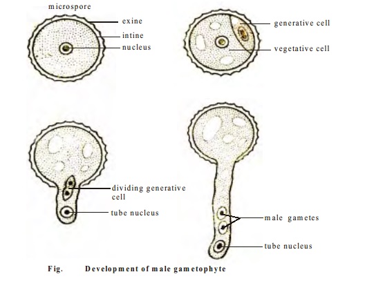
Each pollengrain is unicellular and uninucleate having a two layered wall. The outer layer is called exine and the inner wall is called intine. The exine is provided with spinous outgrowth or different types of ornamentation. The intine is thin, delicate and made up of cellulose.
The exine is not laid uniformly over the intine. The places where exine is not laid is very thin. These thin points are known as germ pore.

Development of male gametophyte
The microspore is the first cell of the male gametophyte. It has a haploid nucleus. The microspore starts germinating while it is still within the microsporangium or pollen sac.
The nucleus of the microspore divides to form a generative nucleus and a tube nucleus or vegetative nucleus. The cell wall is formed resulting in two unequal cells called generative cell and vegetative cell. The generative cell is lenticular or spindle-shaped. Generally, the microspore is shed in the two-celled condition for pollination.
The two celled pollen grain on the stigma of a flower becomes three celled as a result of the division of the generative cell into two male cells or two male gametes. The pollen grain absorbs water and the intine grows out through a germpore to form a pollen tube, which discharges the two male gametes into the embryosac.

Development of female gametophyte
Megasporophyll
The carpels of angiosperm is known as megasporophyll. It is differentiated into three regions-ovary, style and stigma. The ovary contains ovules or megasporangium.
Megasporangium or ovule
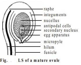
An ovule or megasporangium may arise from the inner surface of the base of an ovary. Each ovule is attached to the placenta by a stalk called funicle. The point of attachment of the ovule to the funicle is known as hilum. The funicle continues beyond the hilum along the body of the ovule and forms a ridge called raphe.
The body of the ovule consists of a parenchymatous mass of tissue called nucellus. The nucellus is surrounded by one or two coverings called integuments. The integuments do not completely cover the nucellus, but leaves a small opening at the tip called micropyle.

Megasporogenesis
Usually a single hypodermal initial known as primary archesporial cell is differentiated at the apex of the nucellus. The primary archesporial cell divides periclinally into outer primary parietal cell or primary wall cell and inner primary sporogenous cell.
The primary parietal cell may or may not divide. The primary sporogenous cell directly behave as megaspore mother cell. The megaspore mother cell undergoes meiotic division to form four megaspores. The four megaspores thus formed are arranged in an axial row forming a linear tetrad. Usually only one megaspore of the tetrad is functional and grows at the expense of other three, which degenerate. The functional megaspore enlarges and forms the embryosac.
Embryosac
The embryosac has three protoplasts of the egg-apparatus towards the micropylar end. Of the three cells constituting the egg-apparatus, one is the egg cell (female gamete) and the other two are known as the synergids. The egg cell, which is enlarged lies below the synergids. At the chalazal end are three antipodal cells. These antipodal cells have no definite function and soon getsdisorganized. In the centre of the embryosac is the secondary nucleus.
Related Topics