Chapter: Human Neuroanatomy(Fundamental and Clinical): The Diencephalon
Ventral Thalamus - The Diencephalon
The Ventral Thalamus
The part of the diencephalon that is called the ventral thalamus lies below the posterior part of the thalamus, behind and lateral to the hypothalamus. In the past it has been referred to as the subthalamic region.
Inferiorly, the ventral thalamus is continuous with the tegmentum of the midbrain (the upper ends of the red nucleus and the substantia nigra reaching it). Laterally, it is related to the lowest part of the internal capsule.
The main masses of grey matter that are included in the ventral thalamus are the reticular nucleus (previously described as part of the dorsal thalamus), the zona incerta and the perigeniculate nuclei.
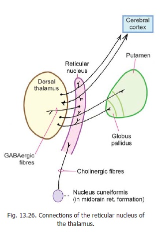
The Reticular Nucleus
The reticular nucleus is made up of a thin layer of neurons covering the lateral aspect of the (dorsal) thalamus, separated from the latter by the external medullary velum. Laterally, the nucleus is related to the internal capsule. Inferiorly, it becomes partially continuous with the zona incerta.
Most fibres emerging from the dorsal thalamus have to traverse the reticular nucleus. (The fibres crossing through it give the nucleus a reticulated appearance, and hence the name). As they pass through it the fibres give collaterals to the reticular nucleus.
In this way the nucleus receives somatic, visceral and auditory impulses. The main efferents of the reticular nucleus pass back into the dorsal thalamus. These fibres are GABAergic. They may influence conduction through the dorsal thalamus (Fig. 13.26). The reticular nucleus also receives fibres from the nucleus cuneiformis (in the reticular formation of the midbrain).
The Pregeniculate Nucleus
This nucleus has connections similar to those of the lateral geniculate nucleus. Its visual connections include fibres from the retina, pretectal region and superior colliculus. It has numerous other connections. The nucleus appears to have some role in vision and in eye movements.
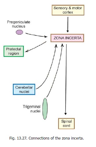
The Zona Incerta
The zona incerta is a thin lamina of grey matter continuous with the reticular nucleus of the thalamus. It intervenes between the subthalamic nucleus and the thalamus. Its functions are not known. Some of its connections are shown in Fig. 13.27.
Advanced:
Lying near the zona inserta there are two groups of neurons that need mention.
1. Some neurons lie along the lower edge of the zona inserta (near the upper end of the red nucleus). These are termed the nuclei of the prerubral field.
2. Some neurons lying within the fibres of the ansa lenticularis constitute the entopeduncular nucleus.
Both these nuclei receive fibres from the globus pallidus and relay them to the reticular formation of the midbrain. Some fibres descend to the inferior olivary complex and to other brainstem nuclei.
Fibre Bundles passing through the Subthalamic Region
In addition to its grey matter the subthalamic region contains a number of fibre bundles (Fig. 13.28,). Ascending tracts (medial lemniscus, spinal lemniscus, trigeminal lemniscus) pass through it on their way from the midbrain to the thalamus. They are accompanied by dentato-thalamic and rubrothalamic fibres.
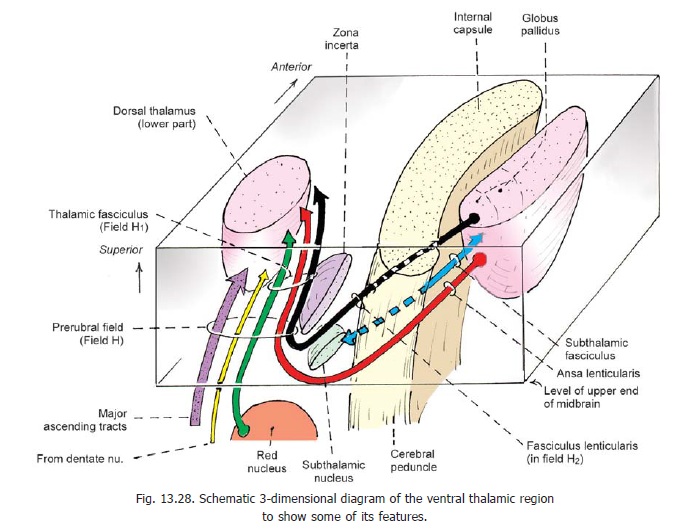
The subthalamic region also contains two bundles of fibres that connect the globus pallidus to the thalamus. These are the ansa lenticularis and the fasciculus lenticularis. Associated with these bundles there are certain regions called the fields of Forel (H, H1, and H2) shown in Fig. 13.28.
Advanced:
Starting from the globus pallidus, the ansa lenticularis winds round the ventral and posterior border of the internal capsule to reach the subthalamic region, where it lies ventral and medial to the subthalamic nucleus. Fibres of the fasciculus lenticularis intersect those of the internal capsule to reach the subthalamic region. Here they pass medially above the subthalamic nucleus and below the zona incerta. This region is field H2 of Forel.
The subthalamic fasciculus (connecting the globus pallidus to the subthalamic nucleus) occupies a position intermediate between the ansa lenticularis and the fasciculus lenticularis. The fibres of the ansa lenticularis and of the fasciculus lenticularis join together medial to the subthalamic nucleus (in field H of Forel) to form thethalamic fasciculus (which is also joined by dentatothalamic and rubrothalamic fibres). The thalamic fasciculus passes above the zona incerta (field H1 of Forel) to reach the thalamus.
Related Topics