Chapter: Human Neuroanatomy(Fundamental and Clinical): The Diencephalon
Medial & Lateral Geniculate Bodies - Thalamus (Dorsal Thalamus)
Medial & Lateral Geniculate Bodies
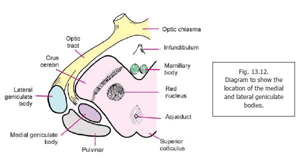
The medial and lateral geniculate bodies are small oval collections of grey matter situated below the posterior part of the thalamus, lateral to the colliculi of the midbrain (Fig. 13.12). Each mass of grey matter is bent on itself, hence the term ‘geniculate’. Traditionally, the geniculate bodies have been grouped together under the heading meta-thalamus; but because of functional relationships they are now included in the dorsal thalamus.
The Medial Geniculate Body
The medial geniculate body is a relay station on the auditory pathway. Medial, ventral and dorsal nuclei are described within it.
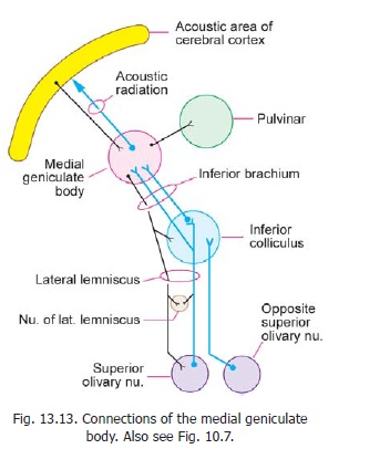
The medial geniculate body receives fibres of the lateral lemniscus either directly, or after relay in the inferior colliculus (Fig. 13.13). These fibres pass through the brachium of the inferior colliculus. Fibres arising in the medial geniculate body constitute the acoustic radiation. The acoustic radiation passes through the sublentiform part of the internal capsule to reach the acoustic areas of the cerebral cortex. Some other connections of the medial geniculate body are shown in Fig. 13.13.
Each medial geniculate body receives impulses from the cochleae of both sides. It also receives fibres from the auditory area of the cerebral cortex. These fibres form part of the descending acousticpathway.
Different neurons in the ventral nucleus of the medial geniculate body respond to different frequencies of sound (tonotopic organisation). The ventral nucleus projects to the primary auditory cortex. The neurons in the dorsal nucleus do not show tonotopic organisation. They project to auditory areas around the primary auditory area.
The Lateral Geniculate Body
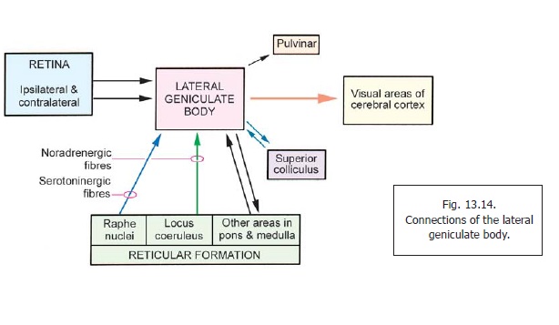
The lateral geniculate body is a relay station on the visual pathway. It receives fibres from the retinae of both eyes (Fig. 13.14). Efferents arising in this body constitute the optic radiation which passes through the retrolentiform part of the internal capsule to reach the visual areas of the cerebral cortex. Details of the visual pathway (including the representation of retinal quadrants in the lateral geniculate body) are described.
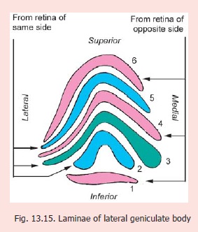
Sections through the lateral geniculate body show that its grey matter is partially split to form six lamellae separated by nerve fibres (Fig. 13.15). These layers are numbered one to six from ventral to dorsal side. Laminae one, four and six receive fibres from the retina of the opposite side; while laminae two, three and five receive fibres from the retina of the same side. The fibres from different parts of the retinae project to specific parts of the lateral geniculate body, the whole of the retina being represented (visiotopic organisation). In turn these project to specific areas of the primary visual cortex (area 17).
Apart from retinal fibres the lateral geniculate body receives fibres from the primary visual cortex, and from extrastriate visual areas. It also receives fibres from the superior colliculus, and from the reticular formation of the pons and medulla.
Noradrenergic fibres reach it from the locus coeruleus, and serotoninergic fibres from raphe nuclei (midbrain).
The various fibres reaching the lateral geniculate body form complicated synapses. The synaptic areas often form glomeruli somewhat similar to those in the cerebellum. In addition to neurons that relay retinal impulses to the visual cortex, the lateral remain confined within it (Golgi type II neurons) and participate in the synapses.
The lateral geniculate body has traditionally been regarded as a simple relay station on the visual pathway. However, both anatomical and physiological evidence shows that considerable interaction of various impulses occurs in the geniculate body. However, the predominant view at present is that impulses from the right and left retinae are not integrated in the lateral geniculate body. Such integration takes place only in the cerebral cortex.
Related Topics