Chapter: 11th 12th std standard Home Science Maintain Basic Knowledge for family life Higher secondary school College
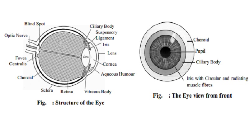
Structure of the Eye
The eye
Structure of the eye
The eye is the organ of the sense of sight situated in the orbital
cavity. It is almost spherical in shape and is about 2.5 cm in diameter. It is
possible to see with only one eye but three - dimensional vision is impaired
when only one eye is used. There are 3 layers of tissue in the wall of the eye.
They are:
The outer fibrous layer : sclera & cornea
The middle vascular layer : choroid, ciliary
body and iris.
The inner nervous tissue layer : retina.
Structures inside the eye ball are the lens, aqueous fluid (humour) and
vitreous body.
Sclera and Cornea
The sclera is the white of the eye and
forms the outermost layer of the eyeball and anteriorly is continuous with the
cornea. The sclera maintains the shape of the eyeball.
The cornea
is a clear transparent membrane. Light rays pass through the cornea to reach
the retina. The cornea is convex anteriorly and refracts or bends light rays to
focus them on to the retina.
Choroid
The choroid is the middle layer and rich in
blood vessels and is a deep chocolate brown in colour.
Ciliary body
This consists of muscle fibres and epithelial
cells. It is located anterior to the choroid. It is attached to the suspensory
ligament which in turn is attached to the capsule enclosing the lens.
Contraction
and relaxation of the ciliary muscle changes the thickness of the lens which
refracts light rays entering the eye to focus them on the retina. The
epithelial cells secrete aqueous fluid into the anterior segment of the eye.
i.e. the space between the lens and the cornea.
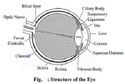
Iris
The iris extends anteriorly from the ciliary body and lies behind the
cornea and in front of the lens. It is a circular body composed of pigment
cells that gives the eye its black, brown, grey or green colour. In the centre
there is an opening called the pupil.
Pupil
varies in size depending upon the intensity of light. In bright light the pupil
constricts. In dim light the pupil dilates.
Lens
The lens is a highly elastic, circular, biconvex, transparent body,
lying immediately behind the pupil. The thickness of the lens is controlled by
the ciliary muscle through the suspensory ligament. It is enclosed within a
transparent capsule.
The lens bends light rays reflected by objects.
Retina
The retina
is the innermost layer of the wall of the eye. It is extremely delicate. It is
composed of several layers of nerve cells and nerve fibres. The retina has a
layer highly sensitive to light, containing cells called rods and cones.
The rods and cones contain photosensitive
pigments that convert light rays into nerve impulses. The rods are for light
vision. The cones are for colour vision.
The rods contain rhodopsin (visual purple) a pigment which is bleached in dim light.
Vitamin A is needed for its synthesis.
Near the centre of the posterior part there is an area which appears
yellow in colour called macula lutea.
In the centre of this area there is a little depression called fovea centralis which consists of only
cone-shaped cells.
About 0.5 cm away from the macula lutea, all the nerve fibres of the
retina converge to form the optic nerve.
The small area of the retina where the optic nerve leaves the eye is the optic disc or
blind spot.
Chambers of the eye
In the anterior segment of the eye, the space
between the cornea and the lens is incompletely divided into anterior and
posterior chambers by the iris. Both chambers contain a clear aqueous fluid (humour) secreted into the chambers by the ciliary glands.
Behind the lens and filling the cavity of the eyeball is the vitreous body (humour). This is a soft,
colourless, transparent, jelly-like
substance composed of 99% water and some salts.
The fluid in both the chambers help to maintain
the shape of the eyeball.
Related Topics