Chapter: 11th 12th standard bio Biotany Plant Tree higher secondary school
Light Microscope and Electron Microscope (TEM - Transmission Electron Microscopy & SEM - Scanning Electron Microscope )
Light Microscope and Electron Microscope (TEM - Transmission Electron Microscopy & SEM - Scanning Electron Microscope )
The modern, complete understanding of cell architecture is based on several types of microscopy. Schleiden and Schwann using a primitive light microscope, first described individual cells as the fundamental unit of life and light microscopy continued to play a major role in biological research. The development of electron microscopes has greatly extended the ability to resolve sub-cellular particles and it has provided new information on the organization of plant and animal tissues. The nature of the image depends on the type of light or electron microscope used and on the way in which the cell or tissue has been prepared for observation.
Light microscopy
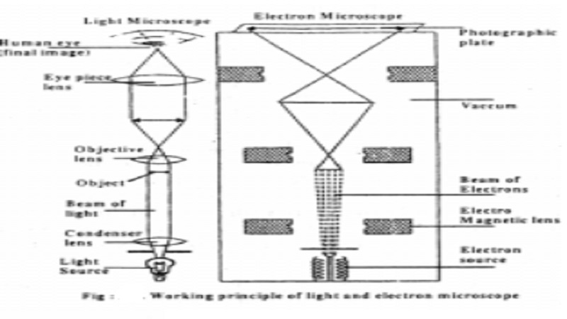
The compound microscope which is most commonly used today contains several lenses that magnify the image of a specimen under study. The total magnification of the object is a product of the magnification of the individual lenses; if the objective lens magnifies 100-fold (a 100 � lens, usually employed) and the eye piece magnifies 10-fold, the final magnification recorded by the human eye or on film will be 1000-fold (100 � 10 ).
The limit of resolution of a light microscope using visible light is about 0.2�m (200nm). No matter how many times the images is magnified, the microscope can never resolve objects that are less than �0.2�m apart or reveal details smaller than �0.2�m in size
Samples for light microscopy are usually fixed, sectioned and stained. Specimens for light microscopy are usually fixed with a solution combining alcohol or formaldehyde, compounds that denature most protein and nucleic acids. Usually the sample is then embedded in paraffin or plastic and cut into sections of one or a few micrometers thick using a microtome. Then these sections are stained using appropriate stains.
Transmission Electron Microscopy

The fundamental principles of electron microscopy are similar to those of light microscopy, the major difference is that in electron microscope electro magnetic lenses and not optical lenses are used. Also it focuses a high velocity electron beam instead of visible light. The electrons are absorbed by atoms in the air and that is the reason why the entire tube between the electron source and the viewing screen is maintained under an ultra high vacuum.
The TEM directs a beam of electrons through a specimen. Electrons are emitted by a tungsten cathode when it is electrically heated. A condenser lens focuses the electron beam on to the sample objective and projects them on to a viewing screen or on a piece of photographic film.
The minimum distance D at which two objects can be distinguished is proportional to the wavelength l of the light that illuminates the objects. Thus the limit of resolution for the electron microscope is theoretically 0.005nm or 40,000 times better than that of unaided human eye. But in reality a resolution of 0.10nm can be obtained with TEM, about 2000 times better than the resolution of light microscopes.
Scanning Electron Microscope
SEM generally has a lower resolving power than the TEM. It is very useful for providing three-dimensional images of the surface of microscopic objects. In this electrons are focused by means of lenses in to a very fine point. The interaction of electrons with the specimen results in the release of different forms of radiation (eg secondary electrons) from the surface of the specimen. These radiations are then captured by an appropriate detector, amplified and then imaged on a television screen.
Other important techniques in EM include the use of ultra thin sections of embedded material; a method of freeze-drying the specimen, which prevents the distortion caused by conventional drying procedure; and the use of negative staining with an electro dense material such as phosphotungstic acid or Uranyl salts. These heavy metal salts provide enough contrast to detect the details of the specimen.
Related Topics