Chapter: Biotechnology Applying the Genetic Revolution: Molecular Biology of Cancer
Detection of Oncogenes by Transformation
DETECTION OF ONCOGENES
BY TRANSFORMATION
If DNA is extracted from cancer
cells and is then inserted into healthy cells, it may change them into cancer
cells. This is known as transformation.
(Note that transformation has a
related but different meaning in bacterial genetics, where it refers to the
uptake and incorporation of any foreign DNA.) Thus, if the presence of an
oncogene is suspected, this can be tested by adding a sample of the suspect DNA
to suitable cells in culture.
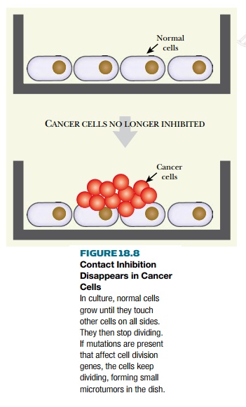
Normal cultured animal cells usually
grow as a thin monolayer on the surface of a culture dish. They do not normally
crawl on top of each other or pile up into heaps (Fig. 18.8). Once the cells on
all sides, they stop dividing, a phenomenon known as contact inhibition. In contrast, cancer cells are uninhibited and
they continue to divide and to aggregate into heaps (see Fig. 18.8). Such
miniature heaps of cells can be seen when DNA containing an oncogene is added
to normal cells in culture. These heaps may be regarded as tiny tumors. If
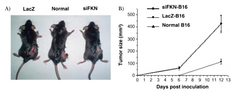
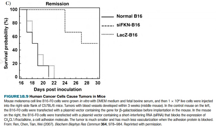
cells from these heaps are injected into an experimental animal such as a
mouse, a real tumor will form (Fig. 18.9).
A related feature of cancer cells is
anchorage independence. Unlike most
normal cells, cancer cells can grow and divide in the absence of binding to
proteins of the extracellular matrix. As a consequence, such cancer cells can
move around and proliferate in “incorrect” locations in the body. This behavior
is especially prominent in malignant (as opposed to benign) tumors.
Related Topics