Chapter: 11th 12th std standard Home Science Maintain Basic Knowledge for family life Higher secondary school College
Blood vessels, Structure and Working of the Heart
Blood vessels
There are several kinds of blood vessels. Arteries and arterioles always carry oxygenated blood from the heart, the exception being the pulmonary arteries which carry venous blood.
Venules and veins carry impure blood towards the heart except the pulmonary veins which carry pure blood.
Capiliaries are very minute blood vessels in which arterioles terminate and venules begin. They form a delicate network of vessels which ramify in most parts of the tissues of the body.
Arteries
They are composed of three layers, tunica adventitia, tunica media and tunica intima. The outer layer is protective in nature. The inner lining is very smooth and lined by a single layer of flat pavement cells. The middle layer is strong. It holds the vessel open and by means of contraction it exerts steady pressure on the blood. The thick walls of the larger arteries are themselves supplied with blood by a special system of tiny vessels known as the vasa-vasorum.
Capillaries
These are microscopic blood vessels through which materials are exchanged between blood and interstitial fluids. They unite to form venules which inturn form veins to carry blood back to heart. Capillaries branch to form an extensive capillary network throughout the tissue. The network increases the surface area allowing a rapid exchange of large quantities of materials. Capillaries are minute vessels in which the arteries terminate. As the arterioles get smaller and smaller, the three coats gradually disappear until when the fine hair-like capillary vessels are formed. These consist of one layer, the inner endothelial coat of the arteries. The extreme thinness of the vessels is highly suitable for filtration, diffusion, osmosis, etc.
Veins
Veins carry blood to heart. Each vein is made up of tunica adventitia, tunica media and tunica intima, similar to that of arteries. But the middle muscular layer is thinner, less firm and elastic than the arteries. At intervals they are thrown out into transverse folds and constitute a sort of incomplete valve. This helps to make the circulation one-way, by allowing blood to flow towards heart but not in opposite direction.
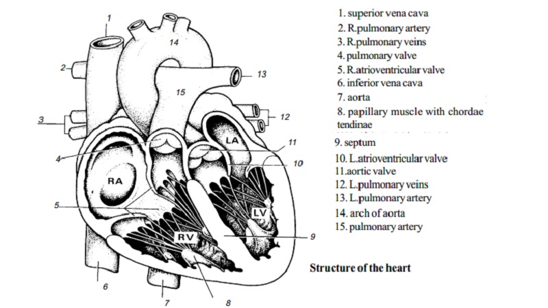
Structure of the Heart
The heart is a cone-shaped, hollow, muscular organ having the base above and the apex below. The apex inclines towards the left
superior vena cava
R.pulmonary artery
R.pulmonary veins
pulmonary valve
R.atrioventricular valve
inferior vena cava
aorta
papillary muscle with chordae tendinae
septum
aortic valve
L.pulmonary artery
arch of aorta
pulmonary artery
side. The heart is about the size of a closed fist. It is divided by a septum into two sides, right and left. Each side is further subdividedinto two chambers, an upper chamber both on the right and the left side is called an atrium or auricle, and a lower chamber, aventricle.
The ventricles have the thickest walls. The walls of the left ventricle is thicker than that of the right as the force of contraction of the left ventricle is much greater. The walls of the auricles are composed of thinner muscles.
The auricles and the ventricles of each side communicate with one another by means of the auriculoventricular opening which are guarded by valves on the right side by the tricuspid vaive and on the left the mitral valve. The auriculoventricular valves permit the passage of blood in one direction only i.e. from auricle to ventricle and they prevent the blood flowing backwards from ventricle to auricle. The interior of each ventricular wall is marked by thickened column of muscle. These project as papillae, the papillary muscles, and at the end attached with thin tendinous cords called the chordae tendineae. They have a second attachment to the lower borders of the auriculoventricular valves and prevents the flaps of the valves being forced up into the auricle when the ventricles contract.
The superior and inferior vena cavae empty their blood into the right auricle. The pulmonary artery carries blood away from the right ventricles to the lungs for purification. The four pulmonary veins bring blood from the lungs to the left ventricle. The openings of the aorta and the pulmonary artery are guarded by the semilunar valves. The valve between the left ventricle and the aorta is called aortic semilunar valve. It prevents blood flowing backwards from the aorta to the left ventricle. The valve between the right ventricle and the pulmonary artery is called pulmonary semilunar valve and prevents blood flowing backwards from the pulmonary artery into the right ventricle.
The heart is composed by a specialised cardiac muscle, surrounded by a membrane of three layers namely the pericardium-outer covering, the myocardium middle muscular layer and the endocardium - the inner lining.
Working of the heart
The heart is a pump and the events which occur in the heart during the circulation of blood are spoken of as the cardiac cycle. In a normal heart beat the two auricles contract simultaneously while the two ventricles relax and vice versa. The term systole refers to the phase of contraction. Diastole is the phase of relaxation. A cardiac cycle consists of the systole and diastole of both auricles and the ventricles.
During the auricular diastole the right auricle receives impure blood from the superior and inferior vena cavae. The left auricle receives pure blood from the pulmonary veins. When the auricle contracts both the auriculoventricular valves are opened and the semilunar valves are closed. So the blood from the right auricle passes into the right ventricle through the tricuspid valve and the pure blood from the left auricle passes into the left ventricle by means of the mitral valve.
In the ventricular systole the ventricles contract and force the blood into their respective vessels. When the ventricles contract the semilunar valves are opened and the auriculoventricular valves are closed. The impure blood from the right ventricle passes through the pulmonary artery to the lungs for purification by opening the pulmonary semilunar valves.
The pure blood from the left ventricle is taken away through the aorta to all over the body by opening the aortic semilunar valve. Thus the blood circulates through the lungs from the right ventricle to the left auricle and through the rest of the body from the left ventricle to the right auricle. The course through the lungs is called pulmonary or lesser circulation, that through all other parts of the body, thesystemic or greater circulation. The average heart beats 72 times/minute. A complete cardiac cycle requires 0.8 seconds.
Pulse
The alternate expansion and elastic recoil of an artery with each systole of the left ventricle is called the pulse. Pulse is strongest in the arteries which are closer to the heart. It becomes weaker as it passes over the arterial system and it disappears altogether in the capillaries. The pulse may be felt in any artery that lies near the surface of the body and over a bone or other firm tissues. The radial artery at the wrist is most commonly used.
The pulse rate is the same as the heart rate and averages between 70 and 90 beats per minute in resting state. If the pulse rate is rapid it is termed as tachycardia. If it is slow, bradycardia. Each pulse beat should be of equal strength. Irregularities in strength may indicate a lack of muscle tone in the heart or arteries.
The Heart Sounds
Heart sounds provide valuable information about the heart valves. The normal heart sounds are usually described by the two syllables !Iubb' and 'dub'. The first sound 'lubb' represents the closing of the auriculoventricular valves. The second sound 'dub' represents the closing of the semilunar valves.
If the sounds are peculiar they are called 'murmurs'. Some murmurs are caused by the noise made by a little blood bubbling back up into an auricle because of improper closure of an auriculoventricular valve. Heart sounds can be easily heard by using stethoscope.
Related Topics