Chapter: Surgical Pathology Dissection : The Digestive System
Appendix: Surgical Pathology Dissection
Appendix
Simple Appendectomies
The
appendix is a common specimen in the surgi-cal pathology laboratory. The
dissection of these specimens is not complex, since most appendec-tomies are
performed for simple acute appendici-tis. Even so, the appendix is all too
often not examined appropriately. Cursory examination of the appendix is a
pitfall to be avoided. Instead, develop the habit of thoroughly examining every
appendix. Regard every appendiceal specimen as an opportunity to uncover
unsuspected patho-logic processes.
The
major objectives in dissecting the simple appendectomy specimen are to document
the presence or absence of inflammation and to search for incidental neoplasms.
These objec-tives are met by examining each component of the appendix—the
serosa, wall, mucosa, and lumen— in a sequential manner. Begin by inspecting
the outer surface of the appendix and the attached mesoappendix. Inflammatory
processes often convert the glistening, smooth, tan serosa into a surface that
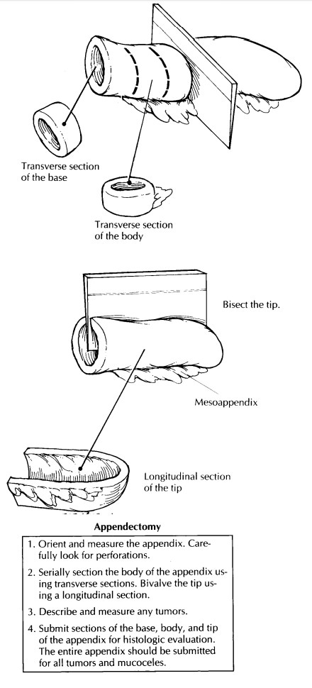
is dull, shaggy, and discolored. Carefully look for perforations. Small
transmural perforations that are not easily seen can some-times be demonstrated
by gently infusing forma-lin into the lumen of the appendix using a syringe. Document
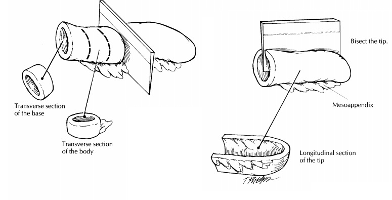
the dimensions of the specimen, and then section the appendix so that the wall,
the mucosa, and the lumen can be evaluated. As illus-trated, bread-loaf the
body of the appendix using thin transverse sections, and bivalve the distal
2-cm tip of the appendix using a longitudinal section. Inspect the wall for
masses, strictures, edema, and other inflammatory changes. Finally, evaluate
the mucosa and the luminal contents for fecaliths, pus, and collections of
mucus. If a neoplasm is present, submit a shave margin from the base of the
appendix, and be sure to document the size of the tumor, the distance from the
tumor to the surgical margin, and the layers of the appendix that are involved.
When the lumen is obstructed, attempt to identify the nature of the
obstruction, keeping in mind that most tumors of the appendix are discovered in
specimens resected for other reasons.
Sections
for histologic evaluation should in-clude a transverse section through the base
and body and a longitudinal section of the tip. Include a portion of the
attached mesoappendix. For a normal-appearing appendix removed by inciden-tal
appendectomy, one section each from the base, body, and tip placed into a
single tissue cas-sette will suffice. For an inflamed appendix, addi-tional sections
may be required to demonstrate points of perforation or luminal obstruction. If
a mass or mucocele is present, the entire appendix should be submitted in a
sequential fashion. The most proximal section from the base of the appen-dix
represents the margin of resection.
Important Issues to Address in Your Surgical Pathology Report on Appendectomies
·
What procedure was performed, and what
structures/organs are present?
·
What are the nature and extent of any
inflam-matory processes present (e.g., acute appendi-citis, abscess formation,
gangrene)? Be sure to mention the presence or absence of perfora-tions and
peritonitis.
·
What are the type, grade, size, location, and
extent of any incidental neoplasms identified? Is the tumor present at the
resection margin?
Related Topics