Chapter: Essentials of Anatomy and Physiology: Digestive System
Anatomy of the oral Cavity
Anatomy of the oral Cavity
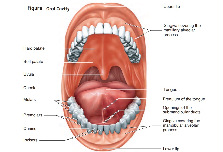
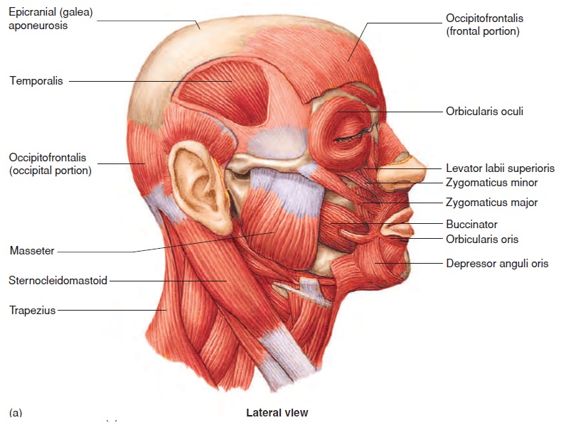
The oral cavity (figure 16.4), or mouth, is the first part of the diges-tive tract. It is bounded by the lips and cheeks and contains the teethand tongue. The lips are muscular structures, formed mostly by the orbicularis oris (̄or-bik′̄u-l̄a′ris̄or′is) muscle (see figure 7.16). Theouter surfaces of the lips are covered by skin. The keratinized strati-fied epithelium of the skin becomes thin at the margin of the lips. The color from the underlying blood vessels can be seen through the thin, transparent epithelium, giving the lips a reddish-pink appearance. At the internal margin of the lips, the epithelium is continuous with the moist stratified squamous epithelium of the mucosa in the oral cavity.
The cheeks form the lateral walls of the oral cavity. The bucci-nator (bŭk′si-nā-tōr) muscles (see figure 7.16), located within thecheeks, flatten the cheeks against the teeth. The lips and cheeks are important in the process of mastication (mas-ti-kā ′ shŭ n), or

They help manipulate the food within the oral cavity and hold the food in place while the teeth crush or tear it. Mastication begins the process of mechanical digestion, which breaks down large food particles into smaller ones. The cheeks also help form words during the speech process.
The tongue is a large, muscular organ that occupies most of the oral cavity. The major attachment of the tongue is in the posterior part of the oral cavity. The anterior part of the tongue is relatively free, except for an anterior attachment to the floor of the mouth by a thin fold of tissue called the frenulum (fren′ ū-lŭm) (figure 16.4). The muscles associated with the tongue are described in chap-ter 7. The anterior two-thirds of the tongue is covered by papillae, some of which contain taste buds . The posterior one-third of the tongue is devoid of papillae and has only a few scattered taste buds. In addition, the posterior portion does contain a large amount of lymphatic tissue, which helps form the lingual tonsil .
The tongue moves food in the mouth and, in cooperation with the lips and cheeks, holds the food in place during mastication. It also plays a major role in the process of swallowing. In addition, the tongue is a major sensory organ for taste, as well as one of the major organs of speech.
Teeth
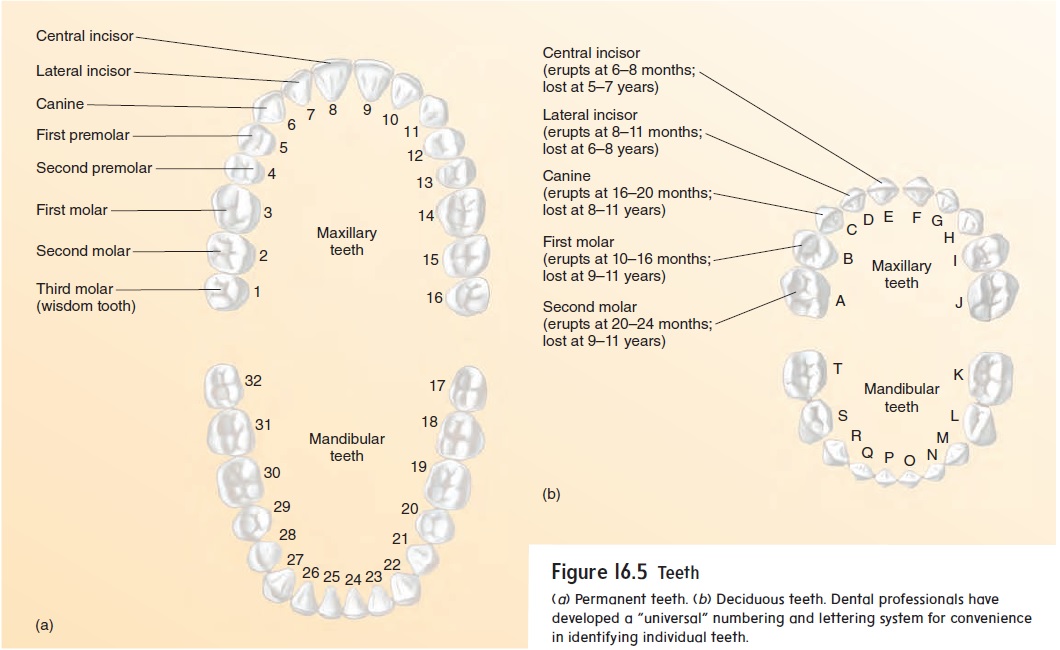
There are 32 teeth in the normal adult mouth, located in the mandible and maxillae. The teeth can be divided into quadrants:right upper, left upper, right lower, and left lower. In adults, each quadrant contains one central and one lateral incisor (in-sı̄′ zŏr; to cut); one canine (kā′ nı̄n; dog); first and second premolars (prē-mō′ lărz; molaris, a millstone); and first, second, and third molars (mō′ lărz). The third molars are called wisdom teeth because they usually appear in the late teens or early twenties, when the person is old enough to have acquired some degree of wisdom.
The teeth of adults are called permanent teeth, or second-ary teeth (figure 16.5a). Most of them are replacements for the20 primary teeth, or deciduous (dē-sid′ ū-ŭs) teeth, also called milk or baby teeth, which are lost during childhood (figure 16.5b).
Each tooth consists of a crown with one or more cusps (points), a neck, and a root (figure 16.6). The center of the tooth is a pulp cavity, which is filled with blood vessels, nerves, and connective tissue, called pulp. The pulp cavity is surrounded by a living, cellular, bonelike tissue called dentin (den′ tin; dens, tooth). The dentin of the tooth crown is covered by an extremely hard, acellular substance called enamel, which protects the tooth against abrasion and acids produced by bacteria in the mouth. The surface of the dentin in the root is covered with cementum (se-men′ tŭm), which helps anchor the tooth in the jaw.
The teeth are rooted within alveoli (al-vē′ ō-lı̄; sockets) along the alveolar processes of the mandible and maxillae. The alveo-lar processes are covered by dense fibrous connective tissue and moist stratified squamous epithelium, referred to as the gingiva (jin′ ji-vă), or gums. The teeth are held in place by periodontal (per′ ē-ō-don′ tăl; around the teeth) ligaments, which are con-nective tissue fibers that extend from the alveolar walls and are embedded into the cementum.
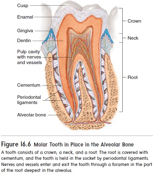
Formation of dental caries (kār′ ēz), or tooth decay, is the result of the breakdown of enamel by acids produced by bacteria on the tooth surface. Enamel is nonliving and cannot repair itself. Consequently, a dental filling is necessary to prevent further dam-age. Periodontal disease is inflammation and degeneration of the periodontal ligaments, gingiva, and alveolar bone. This disease is the most common cause of tooth loss in adults.
Palate and Tonsils
The palate (pal′ ăt), or roof of the oral cavity, separates the oral cavity from the nasal cavity and prevents food from passing into the nasal cavity during chewing and swallowing. The palate consists of two parts. The anterior part contains bone and is called the hardpalate, whereas the posterior portion consists of skeletal muscle andconnective tissue and is called the soft palate (see figure 16.4). The uvula (ū′vū-lă; a grape) is a posterior extension of the soft palate.
The tonsils (ton′ silz) are located in the lateral posterior walls of the oral cavity, in the nasopharynx, and in the posterior surface of the tongue.
Salivary Glands
There are three major pairs of salivary (sal′ i-vār-ē) glands: the parotid, submandibular, and sublingual glands (figure 16.7). A con-siderable number of other salivary glands are scattered throughout the oral cavity, including on the tongue. Salivary glands produce saliva, which is a mixture of serous (watery) and mucous fluids. The salivary glands are compound alveolar glands. They have branching ducts with clusters of alveoli, resembling grapes, at the ends of the ducts .
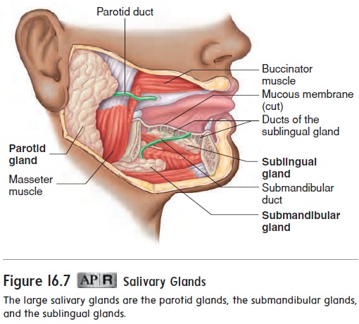
The largest of the salivary glands, the parotid (pă-rot′ id; beside the ear) glands, are serous glands located just anterior to each ear. Parotid ducts enter the oral cavity adjacent to the second upper molars.
Mumps (mŭmpz) is an inflammation of the parotid glandcaused by a viral infection. The inflamed parotid glands become swollen, often making the cheeks quite large. The virus causing mumps can also infect other structures. Mumps in an adult male may involve the testes and can result in sterility.
The submandibular (sŭb-man-dib′ ū-lăr; below the mandible) glands produce more serous than mucous secretions. Each glandcan be felt as a soft lump along the inferior border of the mandible. The submandibular ducts open into the oral cavity on each side of the frenulum of the tongue (see figure 16.4).
The sublingual (sŭb-ling′ gwăl; below the tongue) glands, the smallest of the three paired salivary glands, produce primarily mucous secretions. They lie immediately below the mucous mem-brane in the floor of the oral cavity. Each sublingual gland has 10–12 small ducts opening onto the floor of the oral cavity.
The sublingual (sŭb-ling′ gwăl; below the tongue) glands, the smallest of the three paired salivary glands, produce primarily mucous secretions. They lie immediately below the mucous mem-brane in the floor of the oral cavity. Each sublingual gland has 10–12 small ducts opening onto the floor of the oral cavity.
Related Topics