Chapter: Essentials of Anatomy and Physiology: Digestive System
Anatomy of the large intestine

Anatomy of the large intestine
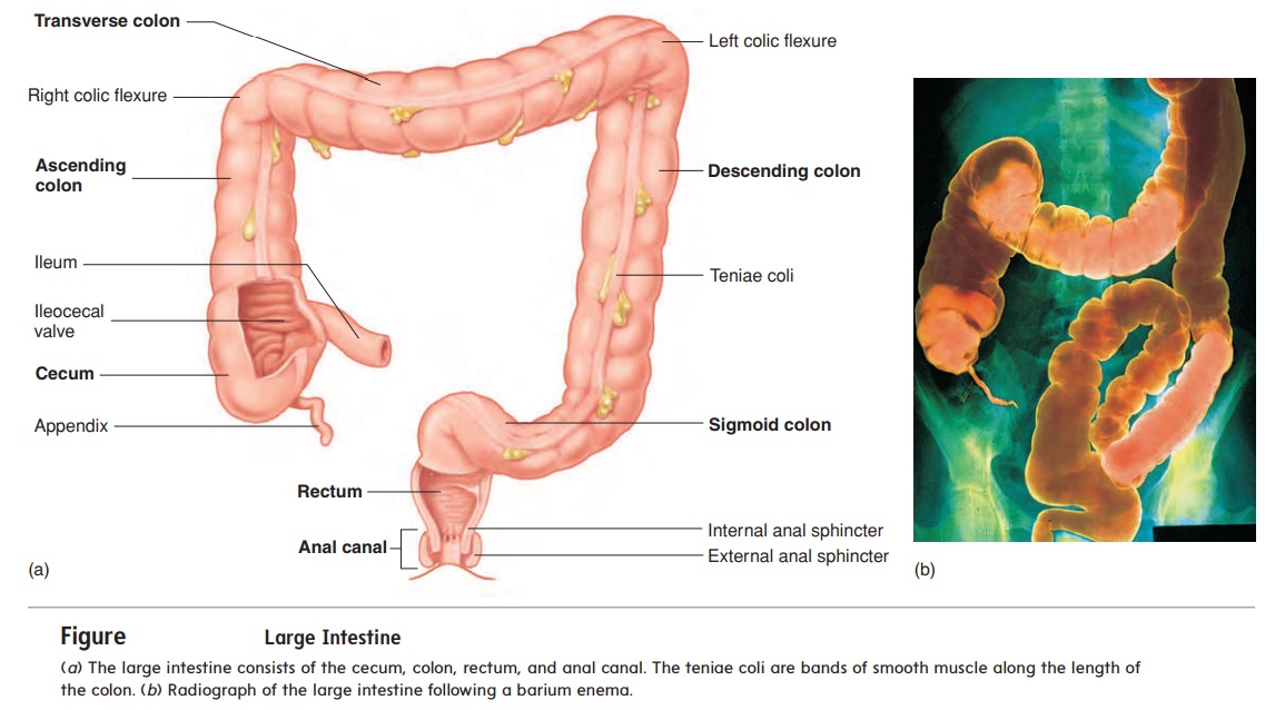
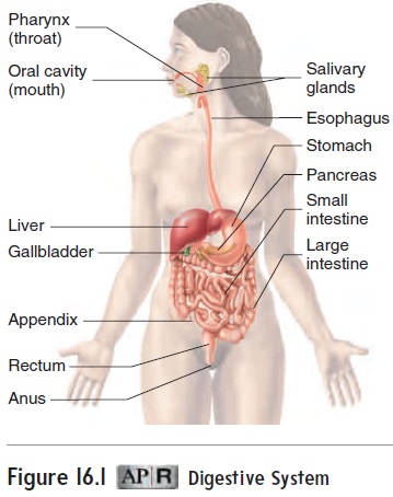
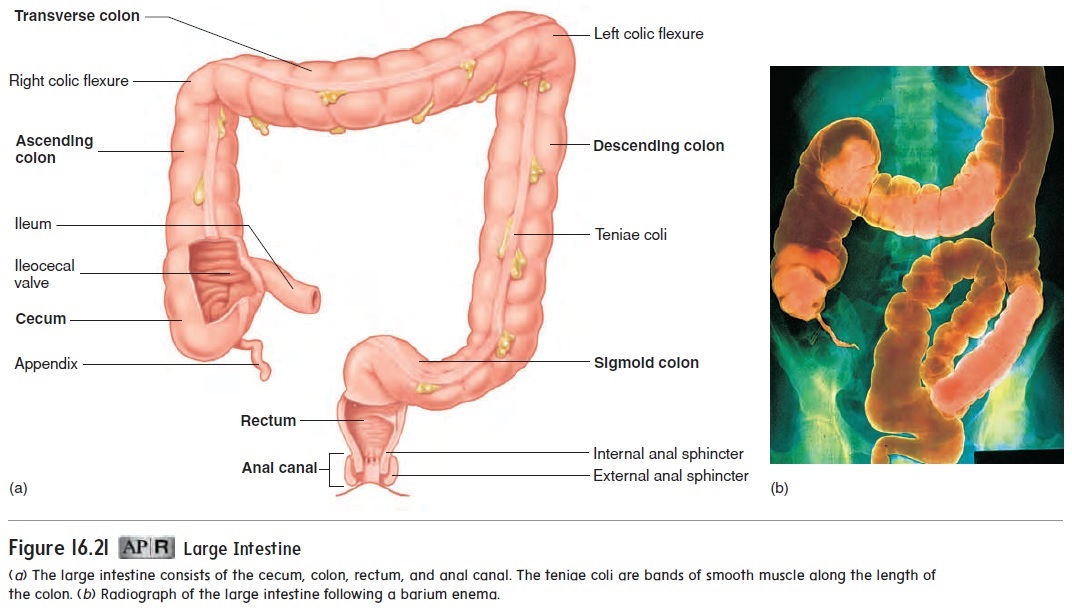
The large intestine consists of the cecum, colon, rectum, and anal canal (figure 16.21; see figure 16.1).


Cecum
The cecum (sē′ kŭm) is the proximal end of the large intestine where it joins with the small intestine at the ileocecal junction. The cecum is located in the right lower quadrant of the abdomen near the iliac fossa. The cecum is a sac that extends inferiorly about 6 cm past the ileocecal junction. Attached to the cecum is a tube about 9 cm long called the appendix.
Colon
The colon (kō′ lon) is about 1.5–1.8 m long and consists of four parts: the ascending colon, the transverse colon, the descending colon, and the sigmoid colon (figure 16.21). The ascending colon extends superiorly from the cecum to the right colic flexure, near the liver, where it turns to the left. The transverse colon extends from the right colic flexure to the left colic flexure near the spleen, where the colon turns inferiorly; and the descending colon extends from the left colic flexure to the pelvis, where it becomes the sigmoid colon. The sigmoid colon forms an S-shaped tube that extends medially and then inferiorly into the pelvic cavity and ends at the rectum.
The mucosal lining of the colon contains numerous straight, tubular glands called crypts, which contain many mucus-producing goblet cells. The longitudinal smooth muscle layer of the colon does not completely envelop the intestinal wall but forms three bands called teniae coli (tē′ nē-ē kō′ l ı̄).
Rectum
The rectum is a straight, muscular tube that begins at the termina-tion of the sigmoid colon and ends at the anal canal (figure 16.21). The muscular tunic is composed of smooth muscle and is relatively thick in the rectum compared to the rest of the digestive tract.
Anal Canal
The last 2–3 cm of the digestive tract is the anal canal. It begins at the inferior end of the rectum and ends at the anus (external diges-tive tract opening). The smooth muscle layer of the anal canal is even thicker than that of the rectum and forms the internal analsphincter at its superior end. The external anal sphincter at theinferior end of the anal canal is formed by skeletal muscle.
Hemorrhoids are enlarged or inflamed rectal, or hemor-rhoidal, veins that supply the anal canal. Hemorrhoids may cause pain, itching, and/or bleeding around the anus. Treatments include increasing bulk (indigestible fiber) in the diet, taking sitz baths, and using hydrocortisone suppositories. Surgery may be necessary if the condition is extreme and does not respond to other treatments.
Related Topics