Chapter: Essentials of Anatomy and Physiology: Digestive System
Anatomy of the liver
Anatomy of the liver
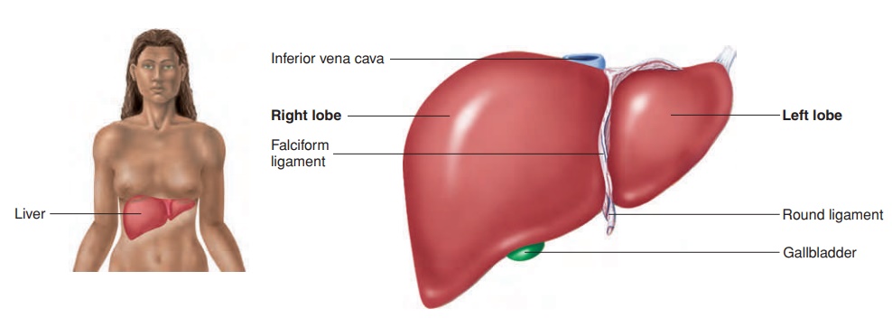
The liver (figure 16.16a,b; see figure 16.1) weighs about 1.36 kilograms (kg) (3 lb) and is located in the right upper quadrant of the abdomen, tucked against the inferior surface of the diaphragm. The posterior surface of the liver is in contact with the right ribs 5–12. It is divided into two major lobes, the right lobe and the left lobe, which are separated by a connective tissue septum, the falciform(fal′si-fōrm) ligament. Two smaller lobes, the caudate (kaw′ dāt; having a tail) lobe and the quadrate (kwah′ drāt; square) lobe, can be seen from an inferior view. Also seen from the infe-rior view is the porta, which is the “gate” through which blood vessels, ducts, and nerves enter or exit the liver.
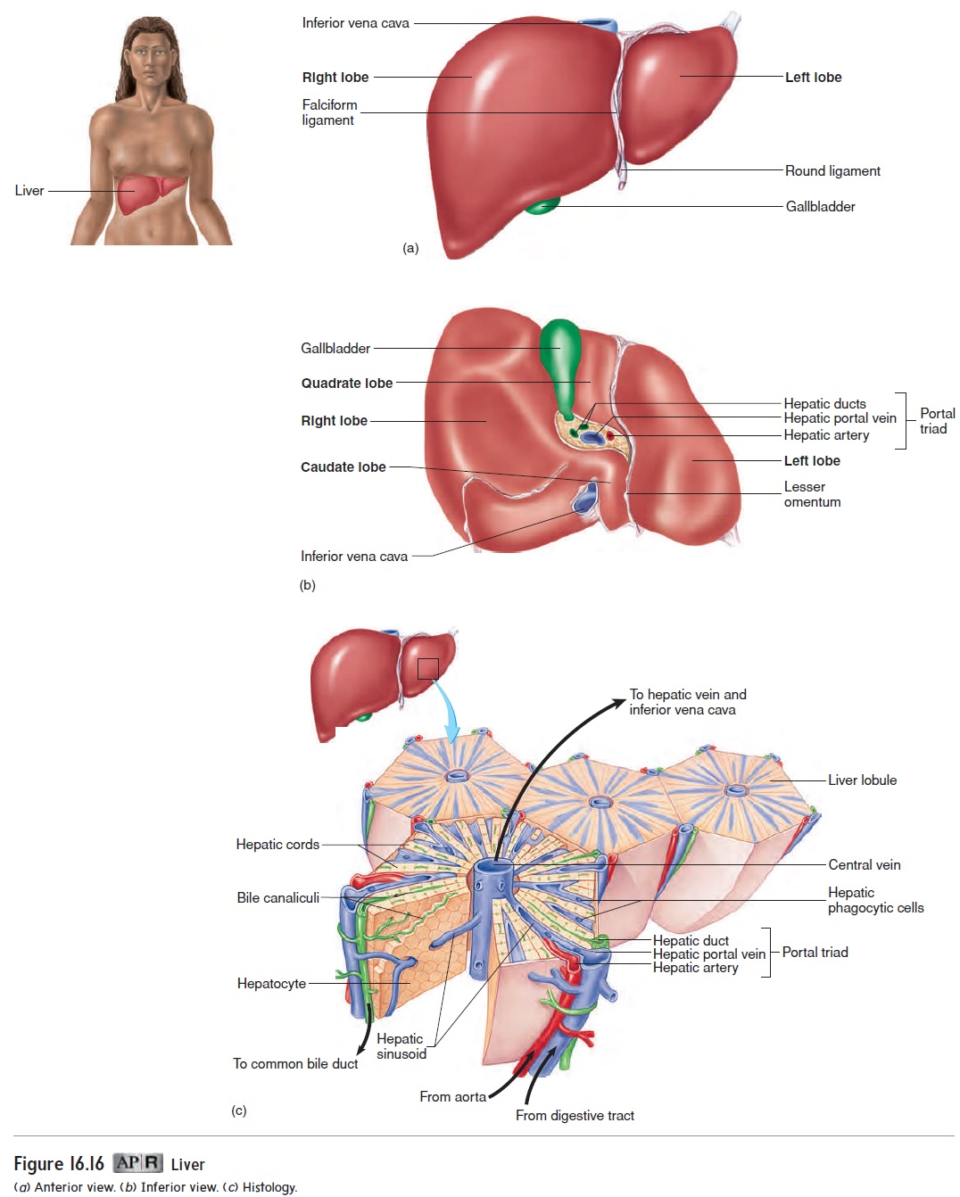
The liver receives blood from two sources . The hepatic (he-pa′ tik; associated with the liver) artery takes oxygen-rich blood to the liver, which supplies liver cells with oxygen. The hepatic portal vein carries blood that is oxygen-poor but rich in absorbed nutrients and other substances from the digestive tract to the liver. Liver cells process nutrients and detoxify harmful substances from the blood. Blood exits the liver through hepatic veins, which empty into the inferior vena cava.
Many delicate connective tissue septa divide the liver into lobules with portal triads at their corners. The portal triads contain three structures: the hepatic artery, the hepatic portal vein, and the hepatic duct (figure 16.16c). Hepatic (he-pa′ tik) cords, formed by platelike groups of cells called hepatocytes (hep′ ă-t ō-sı̄ts), are located between the center and the margins of each lobule. The hepatic cords are separated from one another by blood channels called hepatic sinusoids (si′ nŭ-soydz, sı̄′ nū-soydz; resembling cavities). The sinusoid epithelium contains phagocytic cells that help remove foreign particles from the blood. Blood from the hepatic portal vein and the hepatic artery flows into the sinusoids and mixes together. The mixed blood flows toward the center of each lobule into acentral vein. The central veins from all the lobes unite to form the hepatic veins, which carry blood out of the liver to the inferior vena cava.
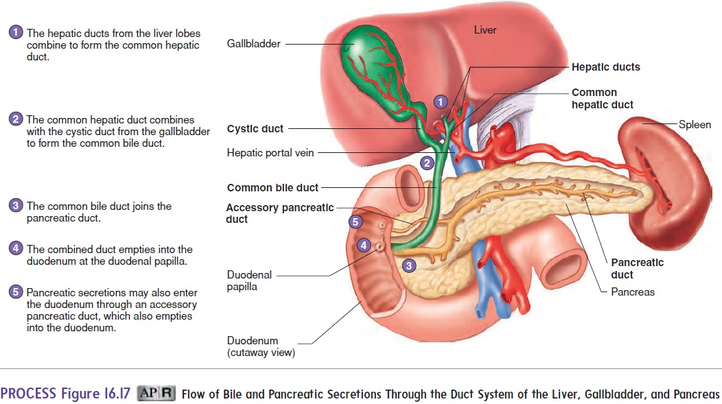
A system of ducts from the liver to the duodenum serves as a pathway for bile and other secretions (figure 16.17). A cleftlike lumen, the bile canaliculus (kan′ ă-lik′ ū-lŭs; pl. kan′ ă lik′ ū-lı̄, little canals), is between the cells of each hepatic cord. Bile, produced by the hepatocytes, flows through the bile canaliculi to the hepatic ducts in the portal triads. The hepatic ducts converge and empty into the right and left hepatic ducts, which transport bile out of the liver. The right and left hepatic ducts unite to form a single common hepatic duct. The common hepatic duct is joined by the cystic (sis′ tik; kystis, bladder) duct from the gallbladder to form thecommon bile duct. The gallbladder is a small sac on the inferior surface of the liver that stores and concentrates bile (see figure 16.16a,b). The common bile duct joins the pancreatic duct and opens into the duodenum at the duodenal papilla (pă-pil′ă) (figure 16.17). The opening into theduodenum is regulated by a sphincter.
Related Topics