Chapter: Essentials of Anatomy and Physiology: Digestive System
Anatomy and Functions of the Pancreas
Anatomy of the Pancreas
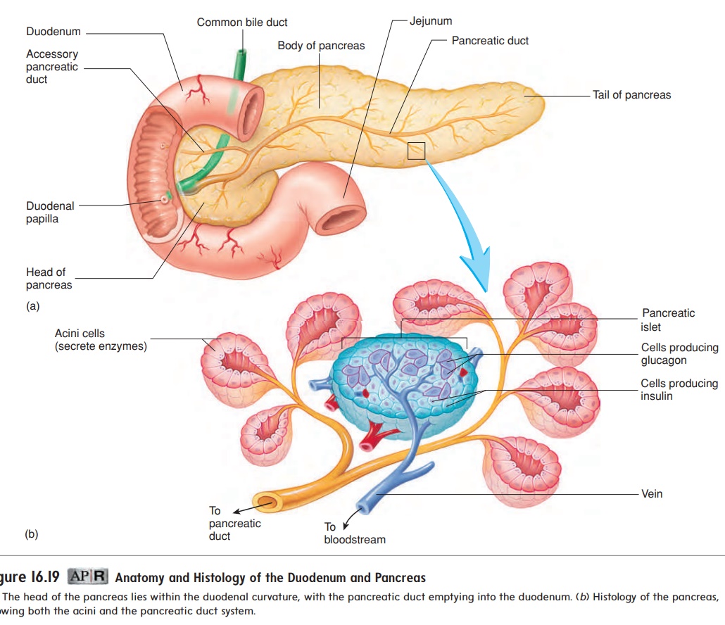
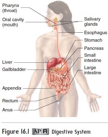
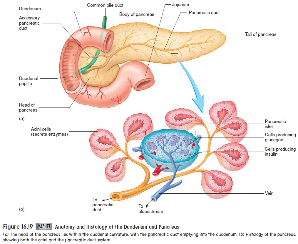
The pancreas is located retroperitoneal, posterior to the stomach in the inferior part of the left upper quadrant (see figure 16.1). It has a head near the midline of the body and a tail that extends to the left, where it touches the spleen (figure 16.19; see figure 16.17). It is a complex organ composed of both endocrine and exocrine tissues that perform several functions. The endocrine part of the pancreas consists of pancreatic islets, or islets of Langerhans. The islet cells produce the hormones insulin and glucagon, which enter the blood. These hormones are very important in controlling blood levels of nutrients, such as glucose and amino acids .
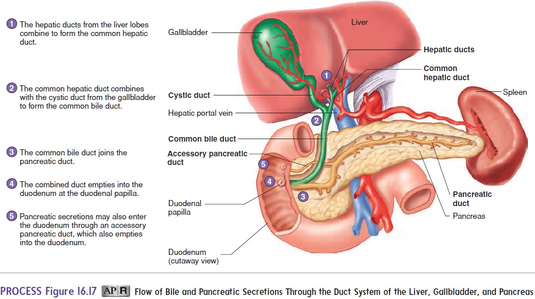
The exocrine part of the pancreas is a compound acinar gland . The acini (as′ i-nı̄; grapes) produce digestive enzymes. Clusters of acini are connected by small ducts, which join to form larger ducts, and the larger ducts join to form the pancreaticduct. The pancreatic duct joins the common bile duct and emptiesinto the duodenum.
Functions Of The Pancreas
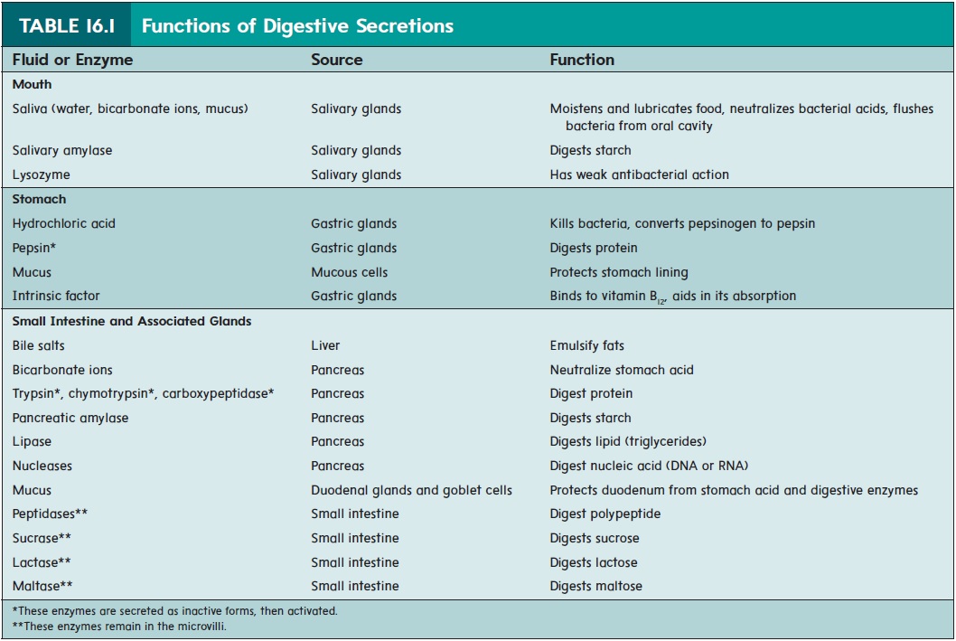
The exocrine secretions of the pancreas include bicarbonate ions (HCO3−), which neutralize the acidic chyme that enters the small intestine from the stomach. The increased pH resulting from the secretion of HCO3− stops pepsin digestion but provides the proper environment for the function of pancreatic enzymes. Pancreatic enzymes are also present in the exocrine secretions and are impor-tant in digesting all major classes of food (see table 16.1). Without the enzymes produced by the pancreas, lipids, proteins, and carbo-hydrates cannot be adequately digested.
The major proteolytic (protein-digesting) enzymes are trypsin(trip′ sin), chymotrypsin (kı̄-mō-trip′ sin), and carboxypeptidase(kar-box′ ē-pep′ ti-dās). These enzymes continue the protein diges-tion that started in the stomach, and pancreatic amylase (am′ il-ās)continues the polysaccharide digestion that began in the oral cavity. The pancreatic enzymes also include lipase (lip′ ās), a lipid-digesting enzyme, andnucleases (noo′ klē-ās-ez), which are enzymes that degrade DNA and RNA to their component nucleotides.
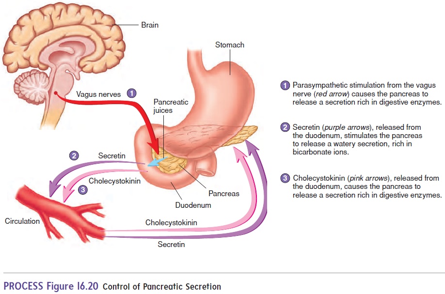
The exocrine secretory activity of the pancreas is controlled by both hormonal and neural mechanisms (figure 16.20; see table 16.2). Secretin initiates the release of a watery pancreatic solution that contains a large amount of HCO3−. The primary stimulus for secretin release is the presence of acidic chyme in the duodenum. Cholecystokinin stimulates the pancreas to release an enzyme-rich solution. The primary stimulus for cholecystokinin release is the presence of fatty acids and amino acids in the duodenum. In turn, enzymes secreted by the pancreas act to digest these fatty acids and amino acids. Parasympathetic stimulation through the vagus nerves also stimulates the secretion of pancreatic juices rich in pancreatic enzymes. Sympathetic action potentials inhibit pancreatic secretion.

Related Topics