Chapter: Clinical Anesthesiology: Regional Anesthesia & Pain Management: Spinal, Epidural & Caudal Blocks
Anatomy of Vertebral Column
Anatomy
THE VERTEBRAL COLUMN
The spine is composed of the vertebral bones
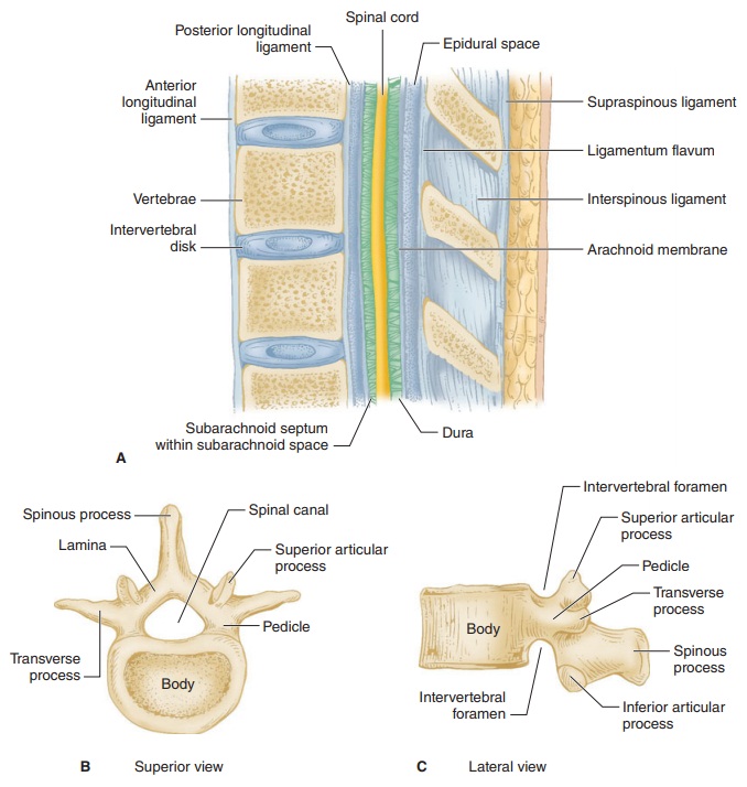
and intervertebral disks (Figure 45–1).
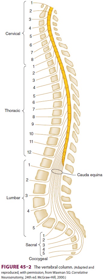
There are 7 cer-vical (C), 12 thoracic (T), and 5 lumbar (L) vertebrae (Figure
45–2). The sacrum is a fusion of 5 sacral (S)
vertebrae, and there are small rudimentary coccy-geal vertebrae. The
spine as a whole provides struc-tural support for the body and protection for
the spinal cord and nerves and allows a degree of mobil-ity in several spatial
planes. At each vertebral level, paired spinal nerves exit the central nervous
system (Figure 45–2).
Vertebrae differ in shape and size at the
various levels. The first cervical vertebra, the atlas, lacks a body and has
unique articulations with the base of the skull and the second vertebra. The
second verte-bra, called the axis, consequently has atypical artic-ulating
surfaces. All 12 thoracic vertebrae articulate with their corresponding rib.
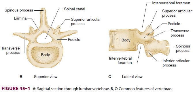
Lumbar vertebrae have a large anterior cylindrical vertebral body. A hollow
ring is defined anteriorly by the vertebral body, lat-erally by the pedicles
and transverse processes, and posteriorly by the lamina and spinous processes
(Figure 45–1B and C). The laminae extend between the transverse processes and
the spinous processes, and the pedicle extends between the vertebral body and
the transverse processes. When stacked verti-cally, the hollow rings become the
spinal canal in which the spinal cord and its coverings sit. The indi-vidual
vertebral bodies are connected by the inter-vertebral disks. There are four
small synovial joints at each vertebra, two articulating with the vertebra
above it and two with the vertebra below. These are the facet joints, which are
adjacent to the transverse processes (Figure 45–1C). The pedicles are notched
superiorly and inferiorly, these notches forming the intervertebral foramina
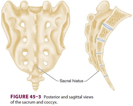
from which the spinal nerves exit. Sacral vertebrae normally fuse into one
large bone, the sacrum, but each one retains discrete anterior and posterior
intervertebral foramina. The laminae of S5 and all or part of S4 normally do
not fuse, leaving a caudal opening to the spinal canal, the sacral hiatus (
Figure 45–3).
The spinal column normally forms a double C, being convex anteriorly in
the cervical and lum-bar regions (Figure 45–2). Ligamentous elements provide
structural support, and, together with supporting muscles, help to maintain the
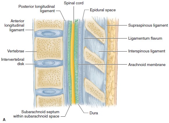
unique shape. Ventrally, the vertebral bodies and inter-vertebral disks are
connected and supported by the anterior and posterior longitudinal ligaments
(Figure 45–1A). Dorsally, the ligamentum flavum,
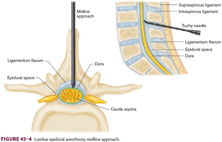
interspinous ligament, and supraspinous ligament provide additional stability.
Using the midline approach, a needle passes through these three dor-sal
ligaments and through an oval space between the bony lamina and spinous
processes of adjacent ver-tebra (Figure 45–4).
Related Topics