Chapter: 11th Zoology : Chapter 4 : Organ and Organ Systems in Animals
Anatomy of Frog : Digestive, Respiratory, Circulatory, Nervous, Excretory, Reproductive system
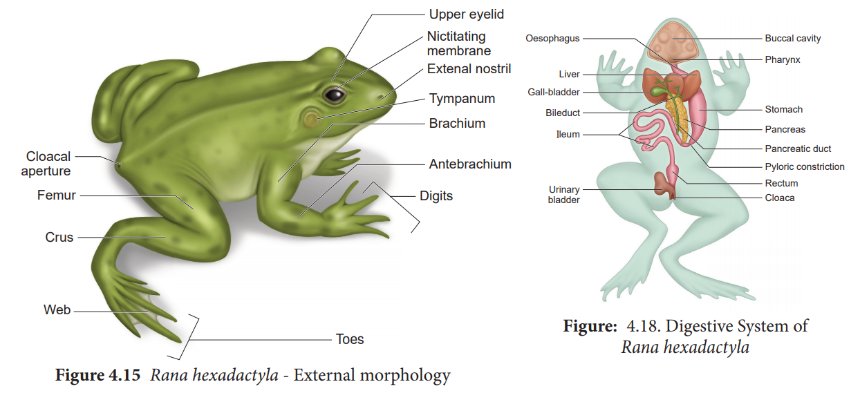
Anatomy
The Digestive System
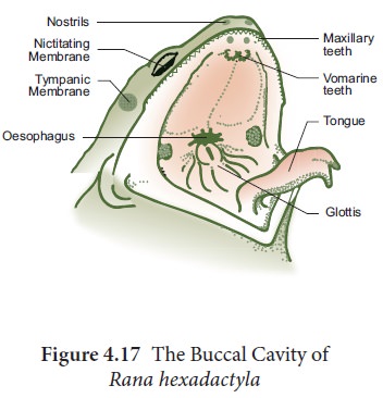
The alimentary canal consists of the buccal cavity, pharynx, oesophagus, duodenum, ileum and the rectum which leads to the cloaca and opens outside by the cloacal aperture. The wide mouth opens into the buccal cavity. On the floor of the buccal cavity lies a large muscular sticky tongue. The tongue is attached in front and free behind. The free edge is forked. When the frog sights an insect it flicks out its tongue and the insect gets glued to the sticky tongue. The tongue is immediately withdrawn and the mouth closes. A row of small and pointed maxillary teeth is found on the inner region of the upper jaw (Figure. 4.17) In addition vomerine teeth are also present as two groups, one on each side of the internal nostrils. The lower jaw is devoid of teeth. The mouth opens into the buccal cavity that leads to the oesophagus through the pharynx. Oesophagus is a short tube that opens into the stomach and continues as the intestine, rectum and finally opens outside by the cloaca
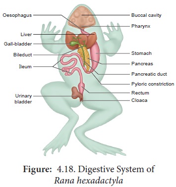
Liver secretes bile which is stored in the gall bladder. Pancreas, a digestive gland produces pancreatic juice containing digestive enzymes.
Food is captured by the bilobed tongue. Digestion of food takes place by the action of Hydrochloric acid and gastric juices secreted from the walls of the stomach. Partially digested food called chyme is passed from the stomach to the first part of the intestine, the duodenum. The duodenum receives bile from the gall bladder and pancreatic juices from the pancreas through a common bile duct. Bile emulsifies fat and pancreatic juices digest carbohydrates, proteins and lipids. Final digestion takes place in the intestine. Digested food is absorbed by the numerous finger-like folds in the inner wall of intestine called villi and microvilli. The undigested solid waste moves into the rectum and passes out through the cloaca.
Respiratory System
Frog respires on land and in the water by two different methods. In water, skin acts
Dissolved oxygen in the water gets, exchanged through the skin by diffusion. On land, the buccal cavity, skin and lungs act as the respiratory organs. In buccal respiration on land, the mouth remains permanently closed while the nostrils remain open. The floor of the buccal cavity is alternately raised and lowered, so air is drawn into and expelled out of the buccal cavity repeatedly through the open nostrils. Respiration by lungs is called pulmonary respiration. The lungs are a pair of elongated, pink coloured sac-like structures present in the upper part of the trunk region (thorax). Air enters through the nostrils into the buccal cavity and then to the lungs. During aestivation and hibernation gaseous exchange takes place through skin.
The Blood-Vascular System
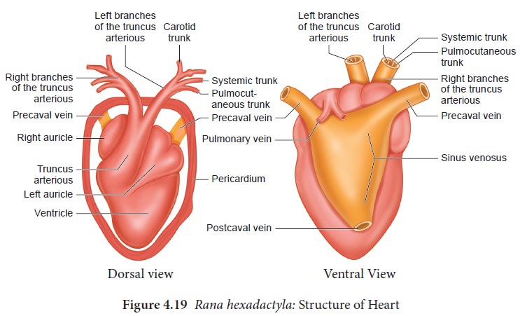
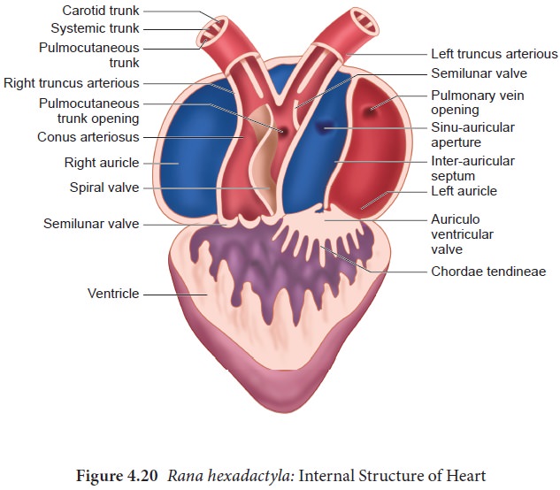
Blood vascular system consists of a heart with three chambers, blood vessels and blood. Heart is covered by a double-walled membrane called pericardium. There are two thin walled anterior chambers called auricles (Atria) and a single thick walled posterior chamber called ventricle. Sinus venosus is a large, thin walled, triangular chamber, which is present on the dorsal side of the heart. Truncus arteriosus is a thick walled and cylindrical structure which is obliquely placed on the ventral surface of the heart. It arises from the ventricle and divides into right and left aortic trunk, which is further divided into three aortic arches namely carotid, systemic and pulmo-cutaneous. The Carotid trunk supplies blood to the anterior region of the body. The Systemic trunk of each side is joined posteriorly to form the dorsal aorta. They supply blood to the posterior part of the body. Pulmo-cutaneous trunk supplies blood to the lungs and skin. Sinus venosus receives the deoxygenated blood from the body parts by two anterior precaval veins and one post caval vein. It delivers the blood to the right auricle; at the same time left auricle receives oxygenated blood through the pulmonary vein. Renal portal and hepatic portal systems are seen in frog (Figure. 4.19 and 420).

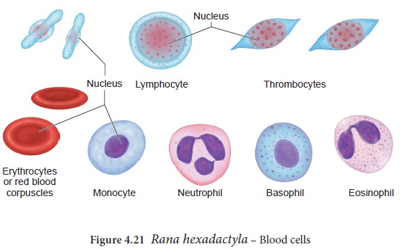
The blood consists of plasma [60%] and blood cells [40 %], red blood cells, white blood cells, and platelets. RBCs are loaded with red pigment, nucleated and oval in shape. Leucocytes are nucleated, and circular in shape (Figure 4.21).
The Nervous System
The Nervous system is divided into the Central Nervous System [CNS], the Peripheral Nervous System [PNS], and the Autonomous Nervous System [ANS]. Peripheral Nervous System consists of 10 pairs of cranial nerves and 10 pairs of spinal nerves. Autonomic Nervous System is divided into sympathetic and parasympathetic nervous system. They control involuntary functions of visceral organs. CNS consists of the Brain and Spinal cord. Brain is situated in the cranial cavity and covered by two meninges called piamater and duramater. The brain is divided into forebrain, midbrain and hindbrain. Fore brain (Prosencephalon) is the anterior most and largest part consisting of a pair of olfactory lobes and cerebral hemisphere (as Telencephalon) and a diencephalon. Anterior part of the olfactory lobes is narrow and free but is fused posteriorly. The olfactory lobes contain a small cavity called olfactory ventricle.
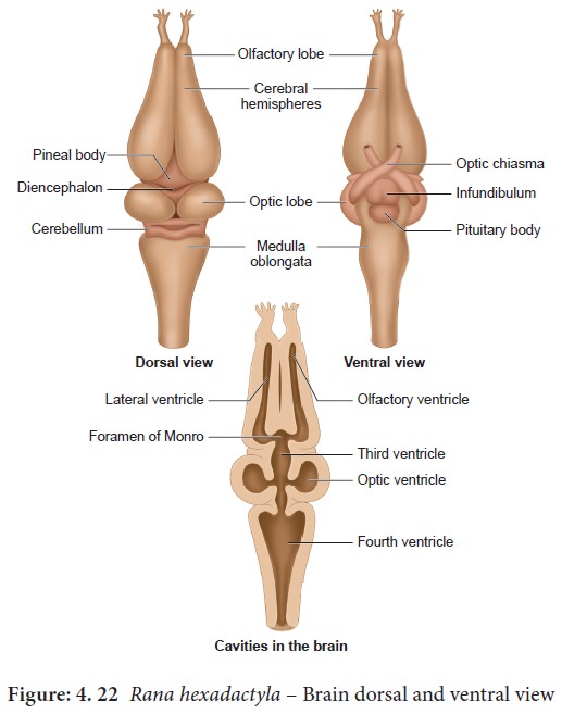
The mid brain (Mesencephalon) includes two large, oval optic lobes and has cavities called optic ventricles. The hind brain (Rhombencephalon) consists of the cerebellum and medulla oblongata. Cerebellum is a narrow, thin transverse band followed by medulla oblongata. The medulla oblongata passes out through the foramen magnum and continues as spinal cord, which is enclosed in the vertebral column (Figure 4.22).
Excretory system
Elimination of nitrogenous waste and salt and water balance are performed by a well developed excretory system. It consists of a pair of kidneys, ureters, urinary bladder and cloaca. Kidneys are dark red, long, flat organs situated on either sides of the vertebral column in the body cavity. Kidneys are Mesonephric. Several nephrons are found in each kidney. They separate nitrogenous waste from the blood and excrete urea, so frogs are called ureotelic organisms. A pair of ureters emerges from the kidneys
Reproductive system
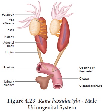
The male frog has a pair of testes which are attached to the kidney and the dorsal body wall by folds of peritonium called mesorchium. Vasa efferentia arise from each testis. They enter the kidneys on both side and open into the bladder canal. Finally, it communicates with the urinogenital duct that comes out of kidneys and opens into the cloaca (Figure 4.23).
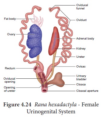
Female reproductive system -(Figure 4.24) consists of paired ovaries, attached to the kidneys, and dorsal body wall by folds of peritoneum called mesovarium. There is a pair of coiled oviducts lying on the sides of the kidney. Each oviduct opens into the body-cavity at the anterior end by a funnel like opening called ostia. Unlike the male frog, the female frog has separate genital ducts distinct from ureters. Posteriorly the oviducts dilated to form ovisacs before they open into cloaca. Ovisacs store the eggs temporarily before they are sent out through the cloaca. Fertilization is external.
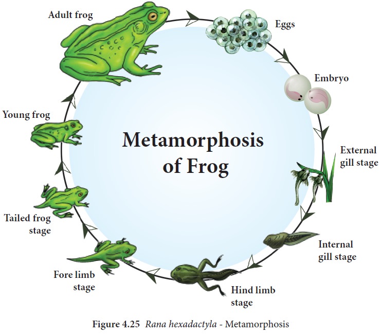
Within few days of fertilization, the eggs hatch into tadpoles. A newly hatched tadpole lives off the yolk stored in its body. It gradually grows larger and develops three pairs of gills. The tadpole grows and metamorphosis into an air – breathing carnivorous adult frog (Figure 4.25). Legs grow from the body, and the tail and gills disappear. The mouth broadens, developing teeth and jaws, and the lungs become functional.
Related Topics