Chapter: 11th Botany : Chapter 9 : Tissue and Tissue System
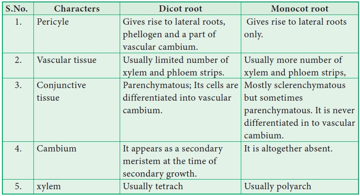
Anatomical differences between dicot root and monocot root
Anatomy of Dicot and Monocot Roots
In
different parts of the plants, the various tissues are distributed in
characteristic patterns. This is best understood by studying their internal
structure by cutting sections (transverse or longitudinal or both) of the part
to be studied.
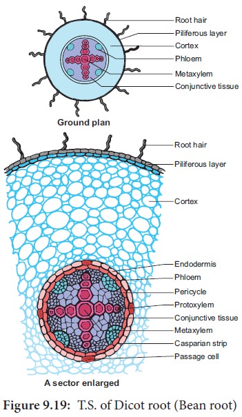
Primary Structure of Dicot Root – Bean Root
The
transverse section of the dicot root (Bean) shows the following plan of
arrangement of tissues from the periphery to the centre.
Piliferous Layer or Epiblema
The
outermost layer of the root is called piliferous
layer or epiblema. It is made up of
single layer of parenchyma cells which are arranged compactly without
intercellular spaces. It is devoid of epidermal pores and cuticle. It possesses
root hairs which are single celled. It absorbs water and mineral salts from the
soil. The chief function of piliferous layer is protection.
Cortex
Cortex
consists of only parenchyma cells. These cells are loosely arranged with
intercellular spaces to make gaseous exchange easier. These cells may store
food reserves. The cells are oval or rounded in shape. Sometimes they are
polygonal due to mutual pressure. Though chloroplasts are absent in the
cortical cells, starch grain are stored in them. The cells also possess
leucoplasts. The innermost layer of the cortex is endodermis. Endodermis is
made up of single layer of barrel shaped parenchymatous cells. Stele is
completely surrounded by endodermis. The radial and the inner tangential walls
of endodermal cells are thickened with suberin
and lignin. This thickening was first noted by Robert Casparay in 1965.
So these thickenings are called casparian strips. But these casparian
strips are absent in the endodermis cells which are located opposite the
protoxylem elements. These thin-walled cells without casparian strips are
called passage cells through which
water and mineral salts are conducted from the cortex to the xylem elements.
Water cannot pass through other endodermal cells due to the presence of
casparian thickenings.
Stele
All the
tissues present inside endodermis comprise the stele. It includes pericycle and
vascular system.
Pericycle
Pericycle
is generally a single layer of parenchymatous cells found inner to the endodermis.
It is the outermost layer of the stele. Lateral roots originate from the
pericycle. Thus, the lateral roots are endogenous in origin.
Vascular System
Vascular tissues are in radial arrangement. The tissue by which xylem and phloem are separated is called conjunctive tissue. In bean, the conjuctive tissue is composed of parenchyma tissue. Xylem is in exarch condition. The number of protoxylem points is four and so the xylem is called tetrach. Each phloem patch consists of sieve tubes, companion cells and phloem parenchyma. Metaxylem vessels are generally polygonal in shape. But in monocot roots they are circular.
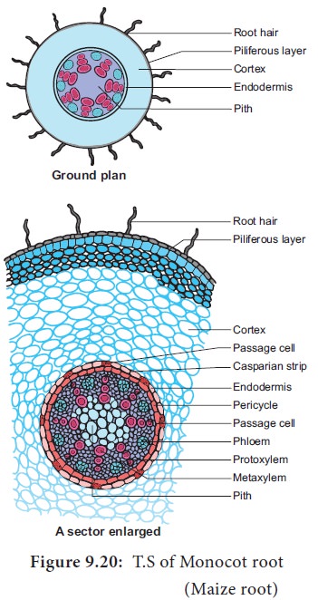
Primary Structure of Monocot Root-maize Root
The transverse section of the monocot root (maize) shows the following plan of arrangement of tissues from the periphery to the centre.
Piliferous Layer or Epiblema
The
outermost layer of the root is known as piliferous
layer. It consists of a single row of
thin-walled parenchymatous cells without any intercellular space. Epidermal
pores and cuticle are absent in the piliferous layer. Root hairs that are found
in the piliferous layers are always unicellular. They absorb water and mineral
salts from the soil. Root hairs are generally short lived. The main function of
piliferous layer is protection of the inner tissues.
Cortex
The
cortex is homogenous. i.e. the cortex is made up of only one type of tissue
called parenchyma. It consists of many layers of thin-walled parenchyma cells
with lot of intercellular spaces. The function of cortical cells is storage.
Cortical cells are generally oval or rounded in shape. Chloroplasts are absent
in the cortical cells, but they store starch. The cells are living and possess leucoplasts. The inner layer of the cortex
is endodermis. It is composed of single layer of barrel shaped parenchymatous
cells. This forms a complete ring around the stele. There is a band like
structure made of suberin and lignin present in the radial and inner
tangential walls of the endodermal cells. They are called casparian strips named after casparay
who first noted the strips. The endodermal cells, which are opposite the
protoxylem elements, are thin walled without casparian strips. These cells are
called passage cells. Their function is to transport water and dissolved salts
from the cortex to the xylem. Water cannot pass through other endodermal cells
due to casparian strips. The main function of casparian strips in the
endodermal cells is to prevent the re -entry of water into the cortex once
water entered the xylem tissue.
Stele
All the
tissues inside the endodermis comprise the stele. This includes pericycle,
vascular system and pith.
Pericycle
Pericycle
is the outermost layer of the stele and lies inner to the endodermis. It consists
of single layer of parenchymatous cells.
Vascular System
Vascular
tissues are seen in radial arrangement. The number of protoxylem groups is
many. This arrangement of xylem is called polyarch. Xylem is in exarch
condition, the tissue which is present between the xylem and the phloem, is
called conjunctive tissue. In maize, the conjunctive tissue is made up of
sclerenchymatous tissue.
Pith
The
central portion is occupied by a large pith. It consists of thin- walled
parenchyma cells with intercellular spaces. These cells are filled with
abundant starch grains.
Related Topics