Chapter: Plant Biochemistry: A leaf cell consists of several metabolic compartments
A leaf cell consists of several metabolic compartments
A leaf cell consists of several metabolic compartments
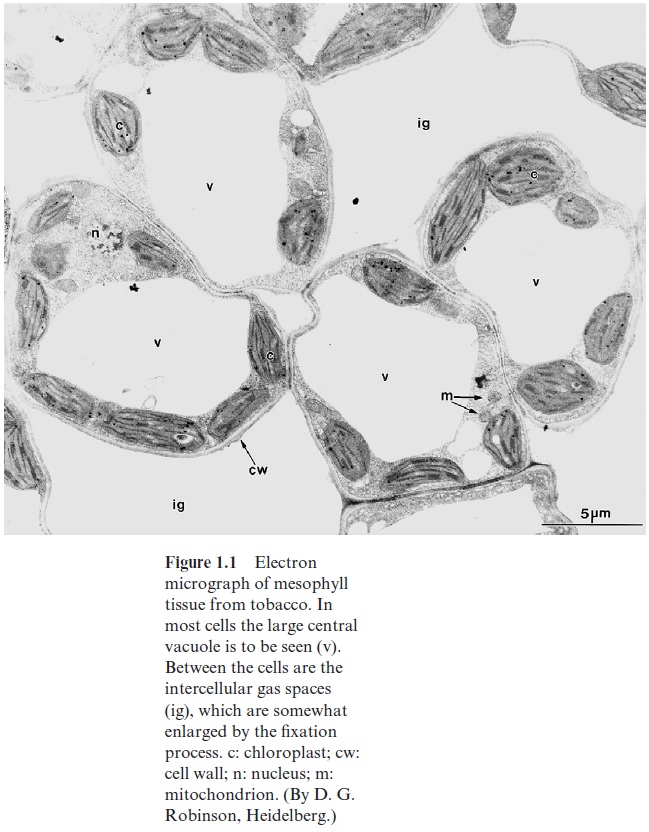
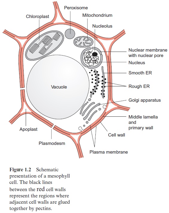
In higher plants photosynthesis occurs mainly in the mesophyll, the chloroplast-rich tissue of leaves. Figure 1.1 shows an electron micrograph of a mesophyll cell and Figure 1.2 shows a schematic presentation of the cell structure. The cellular contents are surrounded by a plasma membrane called the plasmalemma and are enclosed by a cell wall. The cell contains organelles, each with its own characteristic shape, which divide the cell into various compartments (subcellular compartments). Each compartment has specialized metabolic functions. The largest organelle, the vacuole, usually fills about 80% of the total cell volume. Chloroplasts represent the next largest compartment, and the rest of the cell volume is filled with mitochondria, peroxisomes, the nucleus, the endoplasmic reticulum, the Golgi bodies, and, outside these organelles, the cell plasma, called cytosol. In addition, there are oil bodies derived from the endoplasmic reticulum. These oil bod-ies, which occur in seeds and some other tissues (e.g., root nodules), are storage organelles for triglycerides.

The nucleus is surrounded by the nuclear envelope, which consists of the two membranes of the endoplasmic reticulum. The space between the two membranes is known as the perinuclear space. The nuclear envelope is interrupted by nuclear pores with a diameter of about 50 nm. The nucleus contains chromatin, consisting of DNA double strands that are stabilized by being bound to basic proteins (histones). The genes of the nucleus are collectively referred to as the nuclear genome. Within the nucleus, usually off-center, lies the nucleolus, where ribosomal subunits are formed. These ribosomal subunits and the messenger RNA formed by transcription of the DNA in the nucleus migrate through the nuclear pores to the ribosomes in the cytosol, the site of protein biosynthesis. The synthesized proteins are distributed between the different cell compartments according to their final destination

The cell contains in its interior the cytoskeleton, which is a three-dimen-sional network of fiber proteins. Important elements of the cytoskeleton are the microtubuli and the microfilaments, both macromolecules formed by the aggregation of soluble (globular) proteins. Microtubuli are tubular structures composed of α and β tubuline monomers. The microtubuli are con-nected to a large number of different motor proteins that transport bound organelles along the microtubuli at the expense of ATP. Microfilaments are chains of polymerized actin that interact with myosin to achieve movement.
Actin and myosin are the main constituents of the animal muscle. The cytoskeleton has many important cellular functions. It is involved in the spatial organization of the organelles within the cell, enables thermal stabil-ity, plays an important role in cell division, and has a function in cell-to-cell communication.
Related Topics