Chapter: 11th 12th standard bio Biotany Plant Tree higher secondary school
Plant Cell Division - Cell Cycle 1.Amitosis 2.Mitosis 3. Meiosis
Plant Cell Division
Cell Cycle
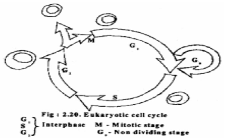
As we have discussed in the earlier chapter, the cell cycle amazingly follows a regular timing mechanism. Most eukaryotic cells live according to an internal clock, that is, they proceed through a sequence of phases, called the cell cycle. During the cell cycle DNA is duplicated during the synthesis(S) phase and the copies are distributed to the daughter cells during mitotic(M) phase. Most growing plant and animal cells take 10-20 hours to double in number and some duplicate at a much slower rate.
A multi cellular organism usually starts it's life as a single cell (zygote). The multiplication of this single cell and it's descendants determine the growth and development of the organism and this is achieved by cell division. Cell division is a complex process by which cellular material is equally divided between daughter cells. Cell division in living things are of three kinds. They are 1.Amitosis 2.Mitosis 3. Meiosis.
Amitosis
It is a simple type of division where the cell contents including nucleus divide into two equal halves by an inwardly growing constriction in the middle of the cell. This type of cell division is common in prokaryotes.
Mitotic cell cycle
It is represented by DNA duplication followed by nuclear division (Karyokinesis) which in turn is followed by cytokinesis. Mitotic cell division was first described by W. Flemming in 1882. In the same year, mitosis in plants was described by Strasburger.
In plants, active mitotic cell division takes place in apices. In higher animals mitotic cell division is said to be diffused, distributed all over the body.
Mitotic cell cycle consists of long interphase(which is sub divided into G1, S and G2 phases), a short M stage (or mitotic stage, subdivided into prophase metaphase, anaphase and telophase) and cytokinesis. The duration of interphase and M-phase varies in different cells.
Interphase
It is the stage in between two successive cell divisions during which the cell prepares itself for the process by synthesizing new nucleic acids and proteins. Chromosomes appear as chromatin network. Interphase consists of the following three sub stages.
i) G1 or Gap-1 phase
This phase starts immediately after cell division. The cell grows in size and there is synthesis of new proteins and RNA needed for various metabolic activities of the cell. A non-dividing cell does not proceed beyond G1 phase. The differentiating cells are said to be in G0 stage.
ii) S-or Synthetic Phase
During this phase there is duplication of DNA. Thus each chromosome now is composedof two sister chromatids.
iii) G2 or Gap-2Phase
The proteins responsible for the formation of spindle fibres are synthesised during this stage.
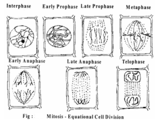
Mitosis
Mitosis is divided into the following 4 sub stages.
1.Prophase 2. Metaphase 3.Anaphase 4. Telophase
1. Prophase
The chromatin network begins to coil and each chromosome becomes distinct as long thread like structure. Each chromosome at this stage has two chromatids that lie side by side and held together by centromere. The nucleus gradually disappears. The nuclear membrane also starts disappearing.
2. Metaphase
The disappearance of nuclear membrane and nucleolus marks the beginning of metaphase.The chromosomes become shorter by further coiling. Finally, the chromosomes become distinct and visible under compound microscope. The chromosomes orient themselves in the equator of the cell in such a way that all the centromeres are arranged in the equator forming metaphase plate or equatorial plate. Out of the two chromatids of each chromosome, one faces one pole and the other one faces the opposite pole. At the same time spindle fibres arising from the opposite poles are seen attached to the centromeres. The fibres are made up of proteins rich in sulphur containing amino acids.
At late metaphase, the centromeres divide and now the chromatids of each chromosome are ready to be separated.
3. Anaphase
Division of centromere marks the beginning of anaphase. The spindle fibres start contracting and this contraction pulls the two groups of chromosomes towards the opposite poles. As the chromosomes move toward opposite poles they assume V or J or I shaped configuration with the centromere proceeding towards the poles with chromosome arms trailing behind. Such variable shapes of the chromosomes are due to the variable position of centromere.
4. Telophase
At the end of anaphase, chromosomes reach the opposite poles and they uncoil, elongate and become thin and invisible. The nuclear membrane and the nucleouls reappear. thus, two daughter nuclei are formed, one at each pole.
Cytokinesis
The division of the cytoplam is called cytokinesis and it follows the nuclear division by the formation of cell wall between the two daughter nuclei. The formation of cell wall begins as a cell plate also known as phragmoplast formed by the aggregation of vesicles produced by Golgi bodies. These vesicles which contain cell wall materials fuse with one another to form cellmembranes and cell walls. Thus, at the end of mitosis, two identical daughter cells are formed.
Significance of Mitosis
As a result of mitosis two daughter cells which are identical to each other and identical to the mother cell are formed.
Mitotic cell division ensures that the daughter cells possess a genetical identity, both quantitatively and qualitatively.
Mitosis forms the basis of continuation of organisms.
Asexual reproduction of lower plants is possible only by mitosis.
Vegetative reproduction in higher plants by grafting, tissue culture method are also a consequence of mitosis.
Mitosis is the common method of multiplication of cells that helps in the growth and development of multi-cellular organism.
Mitosis helps in the regeneration of lost of damaged tissue and in wound healing.
The chromosomal number is maintained constant by mitosis for each species.
Meiosis
Meiosis is a process of cell division of the reproductive cells of both plants and animals in which the diploid number of chromosomes is reduced to haploid.
Meiosis is also known as reduction division(RD) since the number of chromosomes is reduced to half. It takes place only in the reproductive cells during the formation of gametes. Meiosis consists of two complete divisions. As a result of this a diploid cell produces four haploid cells. The two divisions of meiosis are meiosis I or heterotypic division and meiosis II or homotypic division. The first division is meiotic or reductional in which the number of chromosomes is reduced to half and the second division is mitoticor equational.
In all the sexually reproducing organism the chromosome number remains constant generation after generation. During sexual reproduction the two gametes male and female, each having single set oof chromosomes (n) fuse to form a zygote.The zygote thus contains twice as many chromosome as a gamete (n+n=2n). In these two sets of chromosomes one set is derived from the male parent and the other set from the female parent. This is how diploids come to possess tow identical sets of chromosomes called homologous chromosomes Meiosis may take place in the life cycle of a plant during any one of the following events.
At the time of spore formation ie. During the formation of pollen grains in anther and megaspores in ovules.
At the time of gamete formation
At the time of zygote germination.
Each meiotic division cycle is divided into same four stages as in mitosis.
Prophase, Metaphase, Anaphase and Telophase. The name of each stage is followed by I or II depending on which division of cycle is involved.
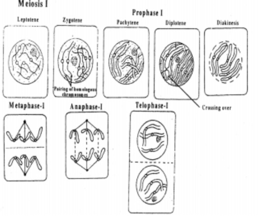
Meiosis I
It consists of four stages namely.
A. Prophase I
B. Metaphase I
C. Anaphase I
D. Telophase I
A. Prophase I
It is the first stage of first meiosis. This is the longest phase of the meiotic division. It includes 5 sub stages namely
1.Leptotene 2.Zygotene 3.Pachytene 4.Diplotene 5.Diakinesis
1. Leptotene
The word leptotene means 'thin thread' . The chromosomes uncoil and become large and thinner. Each chromosome consists of two chromatids.
2. Zygotene
Homologous chromosomes come to-gether and lie side by side throughout their length. This is called pairing or synapsis. The paired chromosomes are now called bivalents. The adjacent non-sister chromatids are joined together at certain posints calledchiasmata.
3. Pachytene
The chromosomes condense further and become very shorter and thicker. They are very distinct now. The two sister chromatids of each homologous chromosome become clearly visible. The bivalent thus becomes a tetrad with four chromatids. In the region of chiasmata, segments of non-sister chromatids of the homologous chromosomes are exchanged and this process is called crossing over
4. Diplotene
The homologous chromosomes condense further. They begin to separate from each other except at the chiasmata. Due to this separation the dual nature of a bivalent becomes apparent and hence the name diplotene.
5. Diakinesis
The Chromosomes continue to contract. The separation of chromosome becomes complete due to terminalisation. The separation starts from the centromeres and goes towards the end and hence the name terminalisation:
The nucleolus and nuclear membrane disappear and spindle formation starts.
B. Metaphase I
The spindle fibres become prominent. The bivalents align on the equatorial plane. Spindle fibres from opposite poles get attached to the centromeres of homologous chromosomes.
C. Anaphase I
The two chromosomes of each bivalent (with chromatids still attached to the centromere) separate from each other and move to the opposite poles of the cell. Thus, only one chromosome of each homologous pair reachers each pole.
Consequently at each pole only half the number of chromosomes (haploid) is received. These chromosomes are, however not the same as existed at the beginning of prophase. Each chromosome consists of one of its original chromatids and the other has a mixture of segments of its own and a segment of chromatid from its homologue (due to crossing over).
D. Telephase I
This is the last stage of meiosis I. Reorganization of the chromosomes at poles occurs to form two haploid nuclei. Nuclear membrane and nucleolus re-appear. The spindle disappears. There is no cytokinesis after meiosis I. The second meiotic division may follow immediately or after a short inter phase. The DNA of the two haploid nuclei does not replicate.
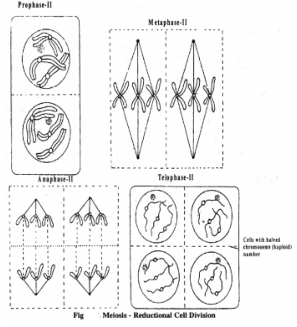
Meiosis II
The second meiotic division is very much similar to mitosis.
Prophase II
The events of prophase II are similar to mitotic prophase. Nucleolus and nuclear membrane disappear. Spindle fibres are formed at each pole.
Metaphase II
Chromosomes move to the centre of the equatorial plane. They get attached to spindle fibres centromere.
Anaphase II
The sister chromatids separate from one another and are pulled to opposite poles of the spindle due to contraction of the spindle fibres.
Telophase II
The chromosomes begin to uncoil and become thin. They reorganize into nucleus with the reappearance of nucleolus and nuclear membrane in each pole. Cytokinesis follows and four haploid daughter cells are formed and thus the meiotic division is completed.
Significance of Meiosis
Meiosis helps to maintain the chromosome number constant in each plant and animal species. In meiosis four haploid daughter cells are formed from a single diploid cell. This is very important in sexual reproduction during the formation of gametes.
The occurrence of crossing over results in the recombination of genes.
The recombination of genes results in genetic variation.
The genetic variations form raw materials for evolution
Related Topics