Chapter: Medical Physiology: Introduction to Physiology: The Cell and Its Functions
Cilia and Ciliary Movements
Cilia and Ciliary Movements
A second type of cellular motion, ciliary movement, is a whiplike movement of cilia on the surfaces of cells.
This occurs in only two places in the human body: on the sufaces of the respiratory airways and on the inside surfaces of the uterine tubes (fallopian tubes) of the reproductive tract. In the nasal cavity and lower respi-ratory airways, the whiplike motion of cilia causes a layer of mucus to move at a rate of about 1 cm/min toward the pharynx, in this way continually clearing these passageways of mucus and particles that have become trapped in the mucus. In the uterine tubes, the cilia cause slow movement of fluid from the ostium of the uterine tube toward the uterus cavity; this move-ment of fluid transports the ovum from the ovary to the uterus.
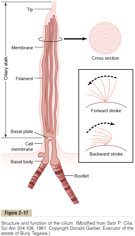
As shown in Figure 2–17, a cilium has the appearance of a sharp-pointed straight or curved hair that projects 2 to 4 micrometers from the surface of the cell. Many cilia often project from a single cell—for instance, as many as 200 cilia on the surface of each epithelial cell inside the respiratory passageways. The cilium is covered by an outcropping of the cell membrane, and it is supported by 11 microtubules—9 double tubules located around the periphery of the cilium, and 2 single tubules down the center, as demonstrated in the cross section shown in Figure 2–17. Each cilium is an out-growth of a structure that lies immediately beneath the cell membrane, called the basal body of the cilium.
The flagellum of a sperm is similar to a cilium; in fact, it has much the same type of structure and same type of contractile mechanism. The flagellum, however, is much longer and moves in quasi-sinusoidal waves instead of whiplike movements.
In the inset of Figure 2–17, movement of the cilium is shown. The cilium moves forward with a sudden, rapid whiplike stroke 10 to 20 times per second, bending sharply where it projects from the surface of the cell. Then it moves backward slowly to its initial position. The rapid forward-thrusting, whiplike movement pushes the fluid lying adjacent to the cell in the direc-tion that the cilium moves; the slow, dragging movement in the backward direction has almost no effect on fluid movement. As a result, the fluid is continually propelled in the direction of the fast-forward stroke. Because most ciliated cells have large numbers of cilia on their sur-faces and because all the cilia are oriented in the same direction, this is an effective means for moving fluids from one part of the surface to another.
Mechanism of Ciliary Movement. Although not all aspects ofciliary movement are clear, we do know the following: First, the nine double tubules and the two single tubules are all linked to one another by a complex of protein cross-linkages; this total complex of tubules and cross-linkages is called the axoneme. Second, even after removal of the membrane and destruction of other ele-ments of the cilium besides the axoneme, the cilium can still beat under appropriate conditions. Third, there are two necessary conditions for continued beating of the axoneme after removal of the other structures of the cilium: (1) the availability of ATP and (2) appropriate ionic conditions, especially appropriate concentrations of magnesium and calcium. Fourth, during forward motion of the cilium, the double tubules on the front edge of the cilium slide outward toward the tip of the cilium, while those on the back edge remain in place. Fifth, multiple protein arms composed of the protein dynein, which has ATPase enzymatic activity, projectfrom each double tubule toward an adjacent double tubule.
Given this basic information, it has been determined that the release of energy from ATP in contact with the ATPase dynein arms causes the heads of these arms to “crawl” rapidly along the surface of the adjacent double tubule. If the front tubules crawl outward while the back tubules remain stationary, this will cause bending.
The way in which cilia contraction is controlled is not understood. The cilia of some genetically abnormal cells do not have the two central single tubules, and these cilia fail to beat. Therefore, it is presumed that some signal, perhaps an electrochemical signal, is transmitted along these two central tubules to activate the dynein arms.
Related Topics