Chapter: Essentials of Anatomy and Physiology: Senses
Vision
VISION
A. List the accessory structures of the eye, and explain their functions.
B. Name the tunics of the eye, list the parts of each tunic, and describe the functions of each part.
C. Explain the differences in function between rods and cones.
D. Describe the chambers of the eye and the fluids they contain.
E. Explain how images are focused on the retina.
The visual system includes the eyes, the accessory structures, and sensory neurons. The eyes are housed within bony cavities called orbits. Action potentials convey visual information from the eyes to the brain. We obtain much of our information about the world through the visual system. For example, education is largely based on visual input and depends on our ability to read words and numbers. Visual input includes information about light and dark, movement and color.
Accessory Structures of the Eye
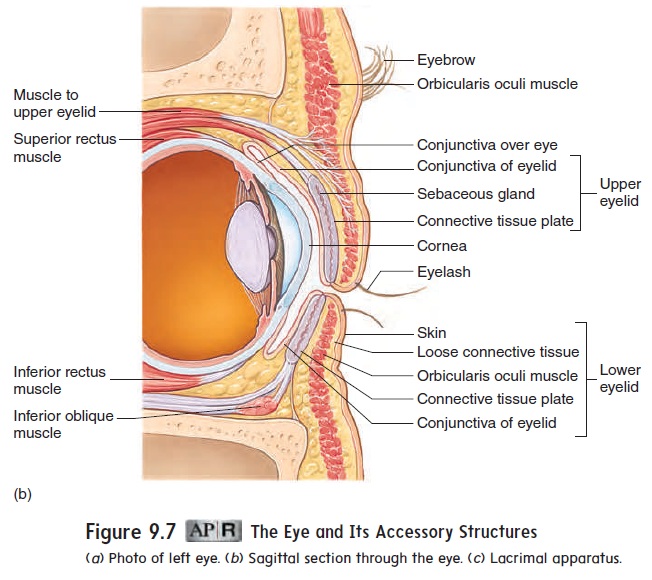
Accessory structures protect, lubricate, and move the eye. They include the eyebrows, eyelids, conjunctiva, lacrimal apparatus, and extrinsic eye muscles (figures 9.7 and 9.8).

Eyebrows
The eyebrows protect the eyes by preventing perspiration from running down the forehead and into the eyes, causing irritation. They also help shade the eyes from direct sunlight (figure 9.7a).
Eyelids
The eyelids, with their associated lashes, protect the eyes from foreign objects (figure 9.7a,b). If an object suddenly approaches the eye, the eyelids protect the eye by closing and then opening quite rapidly (blink reflex). Blinking, which normally occurs about 20 times per minute, also helps keep the eyes lubricated by spreading tears over the surface.
Conjunctiva
The conjunctiva (kon-jŭ nk-t ̄ı ′ v̆a ) is a thin, transparent mucous membrane covering the inner surface of the eyelids and the ante-rior surface of the eye (figure 9.7b). The secretions of the con-junctiva help lubricate the surface of the eye. Conjunctivitis is an inflammation of the conjunctiva (see the Diseases and Disorders table).
Lacrimal Apparatus
The lacrimal (lak′ ri-mă l; tear) apparatus consists of a lacrimal gland situated in the superior lateral corner of the orbit and a nasolacrimal duct and associated structures in the inferior medial corner of the orbit (figure 9.7c). The lacrimal gland produces tears, which pass over the anterior surface of the eye. Most of the fluid produced by the lacrimal glands evaporates from the surface of the eye, but excess tears are collected in the medial angle of the eyes by small ducts called lacrimal canaliculi (kan-̆a -lik′ ̄u -l̄ı ; lit-tle canals). These canaliculi open into a lacrimal sac, an enlarge-ment of the nasolacrimal (n̄a -z̄o -lak′ ri-m̆a l) duct, which opens into the nasal cavity. Tears lubricate and cleanse the eye. They also contain an enzyme that helps combat eye infections.
Extrinsic Eye Muscles
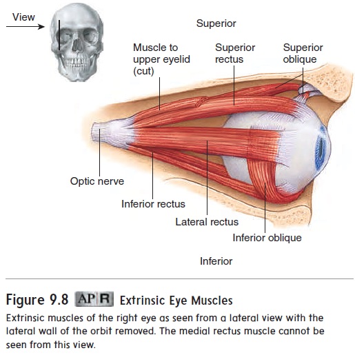
Movement of each eyeball is accomplished by six skeletal muscles called the extrinsic eye muscles (figure 9.8). Four of these muscles run more or less straight from their origins in the posterior portion of the orbit to their insertion sites on the eye, to attach to the four quad-rants of the eyeball. They are the superior, inferior, medial, and lat-eral rectus muscles. Two muscles, the superior and inferiorobliquemuscles, are located at an angle to the long axis of the eyeball.
Anatomy of the Eye
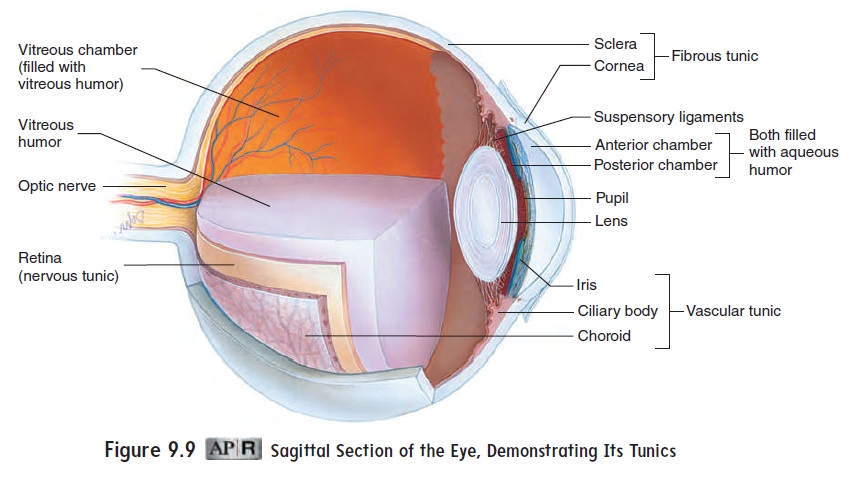
The eyeball is a hollow, fluid-filled sphere. The wall of the eye is composed of three tissue layers, or tunics (figure 9.9). The outer, fibrous tunic consists of the sclera and cornea. The middle, vascular tunicconsists of the choroid, ciliary body, and iris. The inner nervous tunic consists of the retina.
Fibrous Tunic
The sclera (skl̄e r′ ̆a ; hard) is the firm, white, outer connective tis-sue layer of the posterior five-sixths of the fibrous tunic. The sclera helps maintain the shape of the eye, protects the internal structures, and provides attachment sites for the extrinsic eye muscles. A small portion of the sclera can be seen as the “white of the eye.”
The cornea (k̄o r′ n̄e -̆a ) is the transparent anterior sixth of the eye, which permits light to enter. As part of the focusing system of the fibrous tunic, the cornea also bends, or refracts, the entering light.
Vascular Tunic
The middle tunic of the eye is called the vascular tunic because it contains most of the blood vessels of the eye. The posterior por-tion of the vascular tunic, associated with the sclera, is the choroid (k̄o ′ royd). This very thin structure consists of a vascular network and many melanin-containing pigment cells, causing it to appear black. The black color absorbs light, so that it is not reflected inside the eye. If light were reflected inside the eye, the reflection would interfere with vision. The interiors of cameras are black for the same reason.
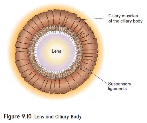
Anteriorly, the vascular tunic consists of the ciliary body and the iris. The ciliary (sil′ ē -ar-ē ) body is continuous with the anterior margin of the choroid. The ciliary body contains smooth muscles calledciliary muscles, which attach to the perimeter of the lens by suspensory ligaments (figure 9.10). The lens is a flex-ible, biconvex, transparent disc (see figure 9.9).
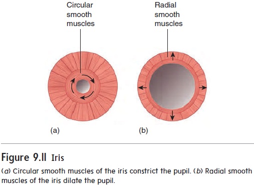
The iris is the colored part of the eye. It is attached to the anterior margin of the ciliary body, anterior to the lens. The iris is a contractile structure consisting mainly of smooth muscle surround-ing an opening called the pupil. Light passes through the pupil, and the iris regulates the diameter of the pupil, which controls the amount of light entering the eye. Parasympathetic stimulation from the oculomotor nerve (III) causes the circular smooth muscles of the iris to contract, constricting the pupil, whereas sympathetic stimula-tion causes radial smooth muscles of the iris to contract, dilating the pupil (figure 9.11). As light intensity increases, the pupil constricts; as light intensity decreases, the pupil dilates.
Nervous Tunic
The nervous tunic is the innermost tunic and consists of the retina. The retina covers the posterior five-sixths of the eye and iscomposed of two layers: an outer pigmented retina and an inner

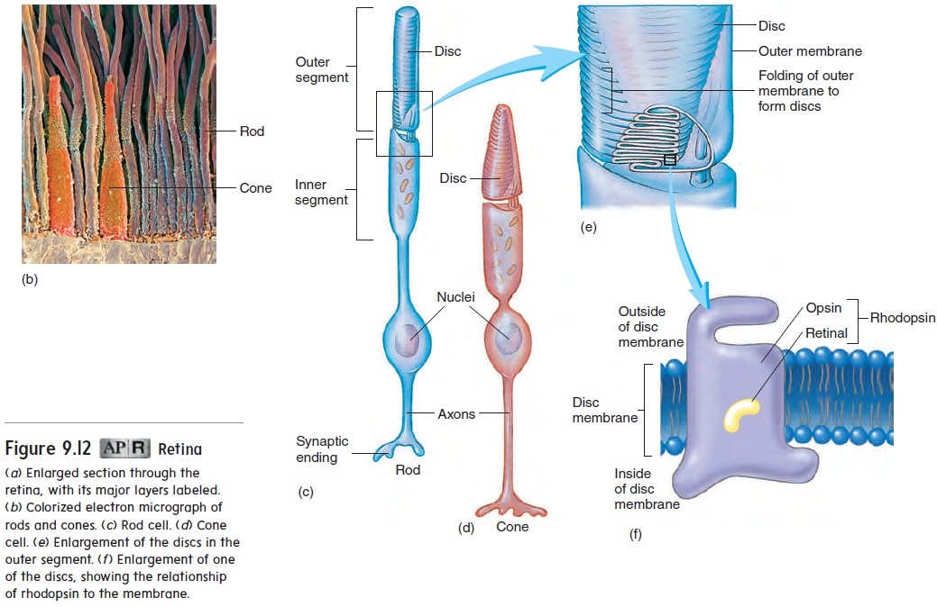
The pigmented retina, with thechoroid, keeps light from reflecting back into the eye. The sen-sory retina contains photoreceptor cells, called rods and cones, which respond to light. The sensory retina also contains numerous interneurons, some of which are named in figure 9.12. Over most of the retina, rods are 20 times more common than cones. Rods are very sensitive to light and can function in dim light, but they do not provide color vision. Cones require much more light, and they do provide color vision. There are three types of cones, each sensitive to a different color: blue, green, or red.
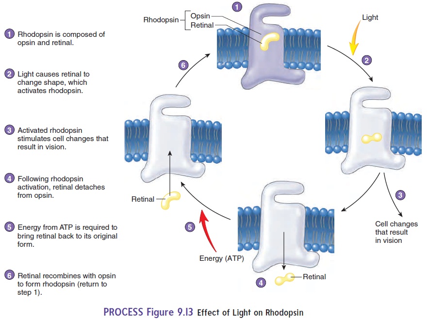
The outer segments of rod and cone cells are modified by numer-ous foldings of the cell membrane to form discs (figure 9.12c–e). Rod cells contain a photosensitive pigment called rhodopsin (rō-dop′ sin; purple pigment). Rhodopsin consists of a protein opsin (op′ sin) loosely bound to a yellow pigment called retinal (ret′ i-năl) (figure 9.12f, figure 9.13, step 1). When exposed to light, retinal changes shape, which then changes the activity of the entire rhodop-sin molecule. This change in rhodopsin stimulates a response in the rod cell, resulting in vision (figure 9.13, steps 2 and 3). Retinal then completely detaches from opsin. Energy (ATP) is required to reat-tach retinal to opsin and return rhodopsin to the form it had before it was stimulated by light (figure 9.13, steps 4–6).
The manufacture of retinal in rod cells takes time and requires vitamin A. In bright light, much of the rhodopsin in rod cells is dissociated (opsin and retinal are separated). For example, suppose you go into a dark building on a bright day. It will take several seconds for your eyes to adjust to the dark as opsin and retinal reassociate to form rhodopsin in the rod cells, which can then react to the dim light. A person with a vitamin A deficiency may have a condition called night blindness, characterized by difficulty seeing in dim light. Night blindness can also result from retinal detachment, which is the separation of the sensory retinafrom the pigmented retina. Retinal detachment affects the periph-ery of the retina, where the rods are located, more than the center of the retina, where the cones are located. Because the rods are more sensitive than the cones to light, retinal detachment affects vision in low light to a greater extent than vision in bright light.
The photosensitive pigments in cone cells are slightly different from those in rod cells. The pigments in cone cells are sensitive to colors. Each color results from stimulation by a certain wavelength of light. Three major types of color-sensitive opsin exist; they are sensi-tive to blue, red, or green. The many colors that we can see result from the stimulation of combinations of these three types of cones.
The rod and cone cells synapse with bipolar cells of the sen-sory retina (see figure 9.12). These and the horizontal cells of the retina modify the output of the rod and cone cells. For example, this modification helps us perceive the borders between objects of contrasting brightness. The bipolar and horizontal cells synapse with ganglion cells, whose axons converge at the posterior of the eye to form the optic nerve(II; see figures 9.9 and 9.12a).
When the posterior region of the retina is examined with an ophthalmoscope (of-thal′ mō-skōp), two major features can be observed: the macula and the optic disc (figure 9.14a). The macula
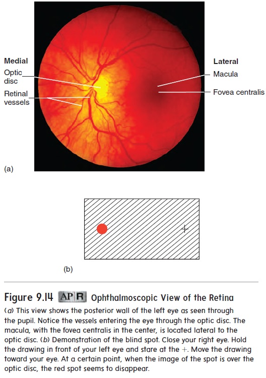
(mak′ ū-lā) is a small spot near the center of the posterior retina. In the center of the macula is a small pit, the fovea (fō′ vē-ă; pit) centralis. The fovea centralis is the part of the retina where lightis most focused when the eye is looking directly at an object. The fovea centralis contains only cone cells, and the cells are more tight-ly packed there than anywhere else in the retina. Hence, the fovea centralis is the region with the greatest ability to discriminate fine images, which explains why objects are best seen straight ahead.
The optic disc is a white spot just medial to the macula, through which a number of blood vessels enter the eye and spread over the surface of the retina. This is also the spot at which axons from the retina meet, pass through the two outer tunics, and exit the eye as the optic nerve. The optic disc contains no photoreceptor cells and does not respond to light; it is therefore called the blindspot of the eye. A small image projected onto the blind spot cannotbe seen (figure 9.14b).
Chambers of the Eye
The interior of the eye is divided into the anterior chamber, the posterior chamber, and the vitreous (vit′rē-ŭs; glassy) chamber(see figure 9.9). The anterior and posterior chambers are located between the cornea and the lens. The iris separates the anterior and the posterior chambers, which are continuous with each other through the pupil. The much larger vitreous chamber is posterior to the lens.
The anterior and posterior chambers are filled with aqueoushumor (watery fluid), which helps maintain pressure within the eye, refracts light, and provides nutrients to the inner surface of the eye. Aqueous humor is produced by the ciliary body as a blood filtrate and is returned to the circulation through a venous ring that surrounds the cornea. The presence of aqueous humor keeps the eye inflated, much like the air in a basketball. If flow of the aqueous humor from the eye through the venous ring is blocked, the pres-sure in the eye increases, resulting in a condition called glaucoma (see the Diseases and Disorders table). Glaucoma can eventually lead to blindness because the fluid com-presses the retina, thereby restricting blood flow through it.
The vitreous chamber of the eye is filled with a transparent, jellylike substance called vitreous humor. The vitreous humor helps maintain pressure within the eye and holds the lens and the retina in place. It also refracts light. Unlike the aqueous humor, the vitreous humor does not circulate.
Functions Of The Eye
The eye functions much like a camera. The iris allows light into the eye, which is focused by the cornea, lens, and humors onto the retina. The light striking the retina produces action potentials that are relayed to the brain.
Light Refraction
An important characteristic of light is that it can be refracted (bent). As light passes from air to some other, denser transparent substance, the light rays are refracted. If the surface of a lens is concave, the light rays are bent, so that they diverge as they pass
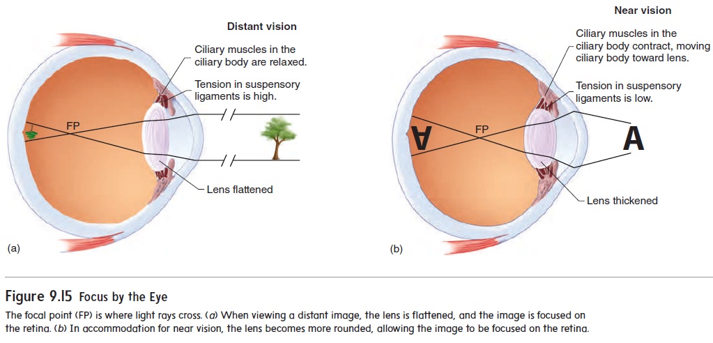
through the lens; if the surface is convex, they converge. As the light rays converge, they finally reach a point at which they cross. The crossing point is called the focal point (FP) (figure 9.15), and causing light to converge is called focusing. The focal point in the eye occurs just anterior to the retina, and the tiny image that is focused on the retina is inverted compared to the actual object.
Focusing Images on the Retina
The cornea is a convex structure, and as light rays pass from the air through the cornea, they converge (figure 9.15). Additional convergence occurs as light passes through the aqueous humor, lens, and vitreous humor. The greatest contrast in media density is between the air and the cornea. The greatest amount of conver-gence therefore occurs at that point. However, the shape of the cornea and its distance from the retina are fixed, so the cornea cannot make any adjustment in focus. Fine adjustments in focus are accomplished by changing the shape of the lens.
When the ciliary muscles are relaxed, the suspensory liga-ments of the ciliary body maintain elastic pressure on the perimeter of the lens, keeping it relatively flat and allowing for distant vision (figure 9.15a). When an object is brought closer than 20 feet (about 6½ m) from the eye, the ciliary muscles contract as a result of parasympathetic stimulation, pulling the ciliary body toward the lens. This reduces the tension on the suspensory ligaments of the lens and allows the lens to assume a more spherical form because of its own internal elastic nature (figure 9.15b). The spherical lens then has a more convex surface, causing greater refraction of light. This process is called accommodation (ă -kom′ ō -dā ′ shŭ n), and it enables the eye to focus on images closer than 20 feet from the eye.
When a person’s vision is tested, a chart is placed 20 feet from the eye, and the person is asked to read a line of letters that has been standardized for normal vision. If the person can read the line, he or she has 20/20 vision, which means that the person can see at 20 feet what people with normal vision see at 20 feet. On the other hand, if the person can only read at 20 feet the line that people with normal vision see at 40 feet, the person’s eyesight is 20/40, and corrective lenses are probably needed.
Neuronal Pathways for Vision
Figure 9.16 shows the neuronal pathways that transmit signals generated by light from the time light enters the eye until it reaches the area of the brain where vision is perceived. The opticnerve leaves the eye and exits the orbit through the optic foramento enter the cranial cavity. Just inside the cranial cavity, the two optic nerves connect to each other at the optic chiasm (k ′ azm; crossing). Axons from the nasal (medial) part of each retina cross through the optic chiasm and project to the opposite side of the brain. Axons from the temporal (lateral) part of each retina pass through the optic nerves and project to the brain on the same side of the body without crossing.
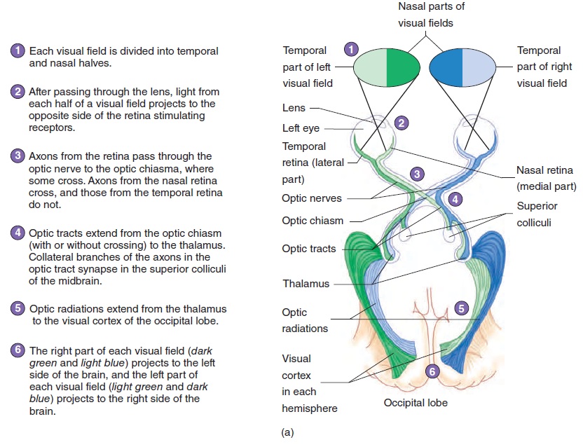
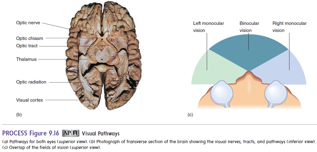
Beyond the optic chiasm, the route of the ganglionic axons is through the two optic tracts (figure 9.16). Most of the optic tract axons terminate in the thalamus. Some axons do not termi-nate in the thalamus but separate from the optic tracts to termi-nate in the superior colliculi, the center for visual reflexes. An example of a visual reflex is turning the head and eyes toward a stimulus, such as a sudden noise or flash of light. Neurons from the thalamus form the fibers of the optic radiations, which project to the visual cortex in the occipital lobe of the cerebrum(figure 9.16). The visual cortex is the area of the cerebrum where vision is perceived.
The image seen by each eye is the visual field of that eye (figure 9.16a). Depth perception (three-dimensional, or binocular, vision) requires both eyes and occurs where the two visual fields overlap (figure 9.16c). Each eye sees a slightly different (mon-ocular) view of the same object. The brain then processes the two images into a three-dimensional view of the object. If only one eye is functioning, the view of the object is flat, much like viewing a photograph.
Related Topics