Chapter: Essentials of Anatomy and Physiology: Senses
Anatomy and function of the Ear
Anatomy and function of the Ear
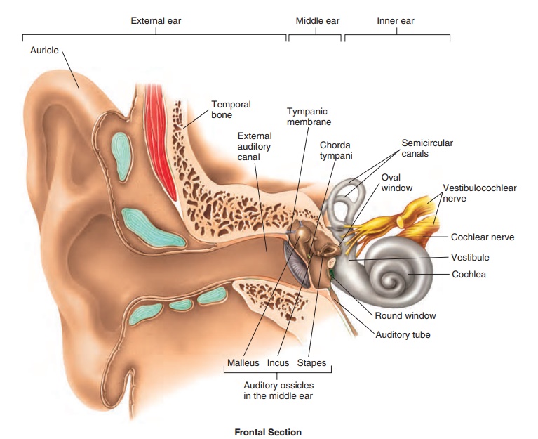
External Ear
The auricle (aw′ ri-kl; ear) is the fleshy part of the external ear on the outside of the head. The auricle opens into the external auditorycanal, a passageway that leads to the eardrum. The auricle collectssound waves and directs them toward the external auditory canal, which transmits them to the tympanic membrane. The auditory canal is lined with hairs and ceruminous (sĕ-roo′ mi-nŭs; cera, wax) glands, which produce cerumen (sĕ-roo′men), a modified sebumcommonly called earwax. The hairs and cerumen help prevent for-eign objects from reaching the delicate tympanic membrane.
The tympanic (tim-pan′ ik; drumlike) membrane, or eardrum, is a thin membrane that separates the external ear from the middle ear. It consists of a thin layer of connective tissue sandwiched between two epithelial layers. Sound waves reaching the tympanic membrane cause it to vibrate.
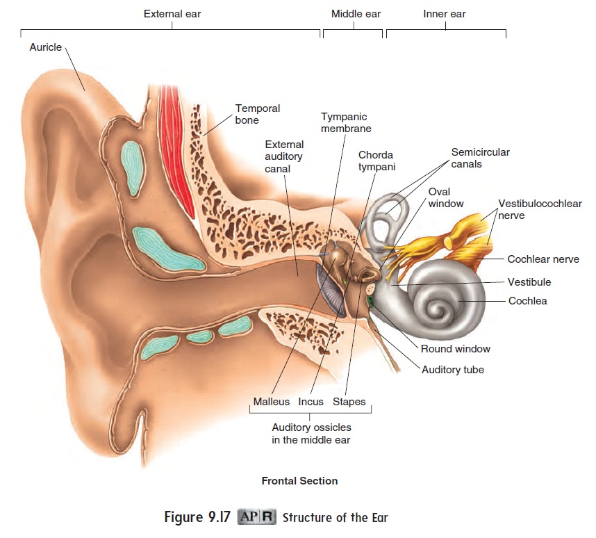
Middle Ear
Medial to the tympanic membrane is the air-filled cavity of the middle ear. Two covered openings on the medial side of the middle ear, the oval window and the round window, connect the middle ear with the inner ear. The middle ear contains three auditoryossicles (os′i-klz; ear bones): the malleus (mal′ē-ŭs; hammer), the incus (ing′kŭs; anvil), and the stapes (stā′pēz; stirrup). These bonestransmit vibrations from the tympanic membrane to the oval win-dow. The malleus is attached to the medial surface of the tympanic membrane. The incus connects the malleus to the stapes. The base of the stapes is seated in the oval window, surrounded by a flexible ligament. As vibrations are transmitted from the malleus to the sta-pes, the force of the vibrations is amplified about 20-fold because the area of the tympanic membrane is about 20 times that of the oval window.
Two small muscles in the middle ear, one attached to the malleus and the other to the stapes, help dampen vibrations caused by loud noises, thus protecting the delicate inner ear structures.
There are two unblocked openings into the middle ear. One opens into the mastoid air cells in the mastoid process of the temporal bone. The other, called the auditory tube, or eustachian (ū-stā′ shŭn) tube, opens into the pharynx and enables air pressure to be equalized between the outside air and the middle ear cavity. Unequal pressure between the middle ear and the outside environ-ment can distort the tympanic membrane, dampen its vibrations, and make hearing difficult. Distortion of the tympanic membrane also stimulates pain receptors associated with that structure. That distortion is why, as a person changes altitude, sounds seem muffled and the tympanic membrane may become painful. These symptoms can be relieved by opening the auditory tube to allow air to enter or exit the middle ear, such as by swallowing, yawning, chewing, or holding the nose and mouth shut while gently forcing air out of the lungs.
Inner Ear
The inner ear consists of interconnecting tunnels and chambers within the temporal bone, called the bony labyrinth (lab′ i-rinth; maze) (figure 9.18a). Inside the bony labyrinth is a smaller set of membranous tunnels and chambers called the membranouslabyrinth (figure 9.18b). The membranous labyrinth is filledwith a clear fluid called endolymph (en′ dō-limf), and the space between the membranous and bony labyrinths is filled with a fluid called perilymph (per′ i-limf). The bony labyrinth can be divided into three regions: the cochlea, the vestibule, and the semicircular canals. The cochlea is involved in hearing. The vestibule and semi-circular canals are involved primarily in balance.
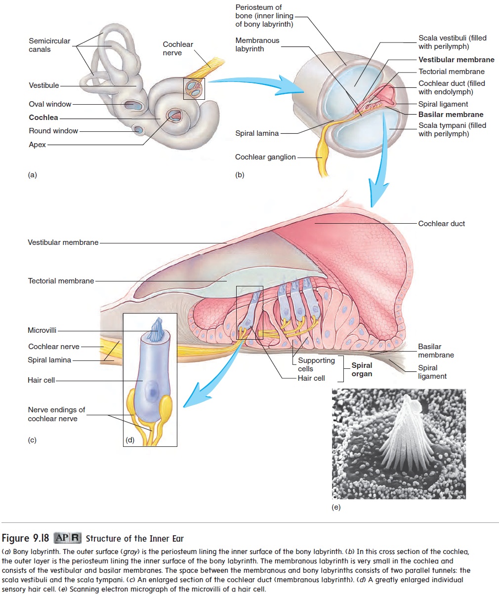
The cochlea (kok′ lē-ă; snail shell) is shaped like a snail shell (figure 9.18a) and contains a bony core shaped like a screw. The threads of this screw are called the spiral lamina. The cochlea is divided into three channels: the scala vestibuli, the scala tympani, and the cochlear duct (figure 9.18b). The scala vestibuli (skā′ lă ves-tib′ ū-l ı̄; scala, stairway) extends from the oval window to the apex of the cochlea. The scala tympani (tim-pa′ nē) extends in parallel with the scala vestibuli from the apex back to the round window. These two channels are perilymph-filled spaces between the walls of the bony and membranous labyrinths. The wall of the membra-nous labyrinth that lines the scala vestibuli is called the vestibular (ves-tib′ ū-lār) membrane; the wall of the membranous labyrinth that lines the scala tympani is the basilar membrane. The cochlearduct is formed by the space between the vestibular membrane andthe basilar membrane and is filled with endolymph.
Inside the cochlear duct is a specialized structure called the spiral organ, ororgan of Corti(figure 9.18c). The spiral organcontains specialized sensory cells called hair cells, which have hairlike microvilli, often referred to as stereocilia, on their sur-faces (figure 9.18c,d,e). The microvilli are stiffened by actin fila-ments. The hair tips are embedded within an acellular gelatinous shelf called the tectorial (tek-tōr′ ē-ăl; a covering) membrane, which is attached to the spiral lamina (figure 9.18b,c).
Hair cells have no axons of their own, but each hair cell is asso-ciated with axon terminals of sensory neurons, the cell bodies of which are located within the cochlear ganglion, or spiral ganglion.
Axons of the sensory neurons join to form the cochlear nerve. This nerve joins the vestibular nerve to become the vestibulocochlearnerve (VIII), which carries action potentials to the brain.
Related Topics