Chapter: Essentials of Anatomy and Physiology: Senses
Functions of the Eye
Functions Of The Eye
The eye functions much like a camera. The iris allows light into the eye, which is focused by the cornea, lens, and humors onto the retina. The light striking the retina produces action potentials that are relayed to the brain.
Light Refraction
An important characteristic of light is that it can be refracted (bent). As light passes from air to some other, denser transparent substance, the light rays are refracted. If the surface of a lens is concave, the light rays are bent, so that they diverge as they pass through the lens; if the surface is convex, they converge.
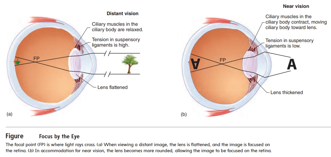
As the light rays converge, they finally reach a point at which they cross. The crossing point is called the focal point (FP) (figure 9.15), and causing light to converge is called focusing. The focal point in the eye occurs just anterior to the retina, and the tiny image that is focused on the retina is inverted compared to the actual object.
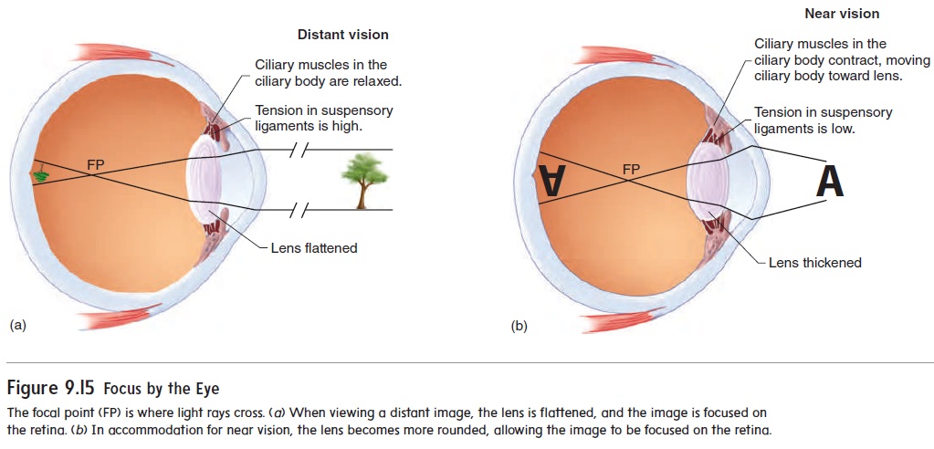
Focusing Images on the Retina
The cornea is a convex structure, and as light rays pass from the air through the cornea, they converge (figure 9.15). Additional convergence occurs as light passes through the aqueous humor, lens, and vitreous humor. The greatest contrast in media density is between the air and the cornea. The greatest amount of conver-gence therefore occurs at that point. However, the shape of the cornea and its distance from the retina are fixed, so the cornea cannot make any adjustment in focus. Fine adjustments in focus are accomplished by changing the shape of the lens.
When the ciliary muscles are relaxed, the suspensory liga-ments of the ciliary body maintain elastic pressure on the perimeter of the lens, keeping it relatively flat and allowing for distant vision (figure 9.15a). When an object is brought closer than 20 feet (about 6½ m) from the eye, the ciliary muscles contract as a result of parasympathetic stimulation, pulling the ciliary body toward the lens. This reduces the tension on the suspensory ligaments of the lens and allows the lens to assume a more spherical form because of its own internal elastic nature (figure 9.15b). The spherical lens then has a more convex surface, causing greater refraction of light. This process is called accommodation (ă -kom′ ō -dā ′ shŭ n), and it enables the eye to focus on images closer than 20 feet from the eye.
When a person’s vision is tested, a chart is placed 20 feet from the eye, and the person is asked to read a line of letters that has been standardized for normal vision. If the person can read the line, he or she has 20/20 vision, which means that the person can see at 20 feet what people with normal vision see at 20 feet. On the other hand, if the person can only read at 20 feet the line that people with normal vision see at 40 feet, the person’s eyesight is 20/40, and corrective lenses are probably needed.
Related Topics