Chapter: Human Nervous System and Sensory Organs : Basic Elements of the Nervous System
The Nerve Cell
The Nerve Cell
The nervous tissue consists of nerve cells and glial cells which originate from the ectoderm (the latter are supporting and covering cells). Blood vessels
and meninges do not belong to the nervous tissue; they are of mesodermal
origin. The nerve cell (gan-glion cell or neuron) is the functional unitof the
nervous system. In its mature state, it is no longer able to divide, thus
making pro-liferation and the replacement of old cells impossible. Very few
nerve cells are formed after birth.
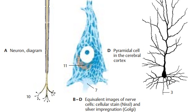
A neuron
consists of the cell body, the peri-karyon
(A1), the processes, dendrites (A2),and one main process, the axon
or neurite (A – D3).
The perikaryon is the trophic center of the cell, and processes that become separated
from it degenerate. It contains the cell
nu-cleus (A4) with a large,
chromatin-rich nucleolus (A5) to which the Barr body (sexchromatin) (A6)
is attached in females.
The dendrites enlarge the cell surface by
branching. The processes of other neurons end here: the dendrites are the sites
where nerve impulses are received. The
processes ofother neurons often end at small dendritic appendices, spines (thorns), which give the
dendrites a rough appearance (D).
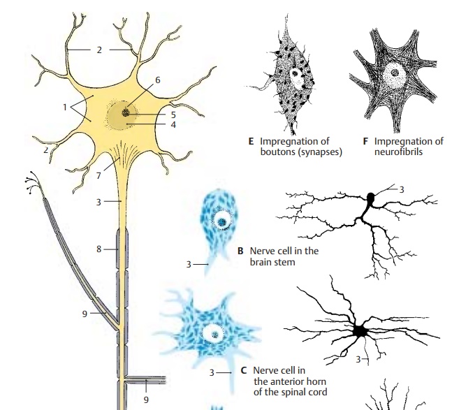
The axon conducts the nerve impulse and
begins with the axon hillock (AD7), the site where nerve impulses are generated. At a
cer-tain distance from the perikaryon (initialsegment)
it becomes covered by themyelinsheath (A8), which consists of a
lipid-con-taining substance (myelin).
The axon gives off branches (axon
collaterals) (A9) and fi-nally
ramifies in the terminal area (A10)
to end with small end-feet (axon
terminals, or boutons) on nerve
cells or muscle cells. Thebouton forms a synapse with the surface membrane of
the next cell in line; it is here that impulse transmission to the other cell
takes place.
Depending
on the number of processes, we distinguish between unipolar, bipolar, or multipolar neurons. Most neurons are
multi-polar. Some have short axons (interneurons), others have axons more than 1 m long (pro-jection neurons).
A neuron
cannot be visualized in its entirety by applying just one staining method. The
different methods yield only partial images of neurons. The cellular stain (Nissl’smethod) shows nucleus and perikaryon (B – D). The latter,
including the bases of the dendrites, is filled with clumps (Nissl sub-stance, tigroid bodies) and may contain pig-ments (melanin, lipofuscin) (D11).
The axon hillock is free of Nissl bodies. The Nissl sub-stance is the
light-microscopic equivalent of a well-developed rough endoplasmatic reti-culum. Motor neurons possess a large
peri-karyon with coarse Nissl bodies, while sensory neurons are smaller and
often con-tain only Nissl granules.
Impregnation with silver (Golgi’s method) stains the entire cell
including allneuronal processes; the cell appears as a brown-black silhouette (B – D). Other im-pregnation methods selectively stain the axon terminals (E), or the neurofibrils (F)running in parallel bundles through
peri-karyon and axon.
Related Topics