Chapter: Human Nervous System and Sensory Organs : Basic Elements of the Nervous System
The Nerve Fiber

The Nerve Fiber
The axon
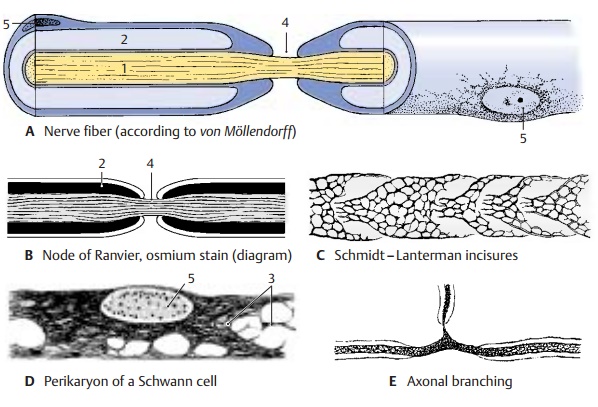
(AFG1) is surrounded by a sheath: in
unmyelinated nerve fibers by the
cyto-plasm of the sheath cells, and in myelinatednerve
fibers by themyelin sheath(ABG2). Axon and sheath together are
called the nerve fiber. The myelin
sheath begins be-hind the initial segment of the axon and ends just before the
terminal ramification. It consists of myelin,
a lipoprotein produced by the sheath cells. The sheath cells in the CNS are oligodendrocytes; in the peripheralnerves
they are Schwann cells, which
origi-nate from the neural crest. The myelin sheath of fresh, unfixed nerve
fibers appears highly refractile and without structure. Its lipid content
makes it birefringent in polarized light. The lipids are removed upon fixation,
and the denatured protein scaffold remains as a gridlike structure (neurokeratin) (D3).

At
regular intervals (1 – 3 mm), the myelin sheath is interrupted by deep
constrictions, the nodes of Ranvier
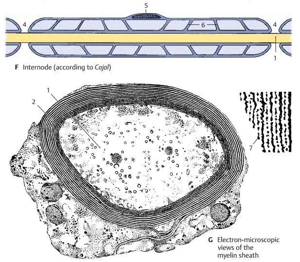
(AB4 F). The segment be-tween two
nodes of Ranvier in peripheral nerves, the internode
or interannular segment (F), corresponds to the expansion of one
sheath cell. The cell nucleus (ADF5)
and perinuclear cytoplasm form a slight bulge on the myelin sheath in the
middle of the in-ternode. Cytoplasm is also contained in ob-lique indentations,
the Schmidt–Lantermanincisures (C, F6) (see also p. 40, A4). The mar-gins
of the sheath cells define the node of Ranvier at which axon collaterals (E) may branch off or synapses may
occur.

Related Topics