Chapter: Human Nervous System and Sensory Organs : Basic Elements of the Nervous System
Neuronal Systems

Neuronal Systems
Groups of neurons that have the
same transmitter and the axons of which often form dense fiber bundles are
described according to their transmitter substance as cholinergic,noradrenergic, dopaminergic, serotoninergic, GABAergic,
or peptidergic systems. The im-pulse
can be transmitted to neurons of the same type or to neurons with a different
neurotransmitter. In the sympathetic nervous system, for example, the neurons
in the spinal cord are cholinergic; however, transmission in the peripheral
ganglia switches to noradrenergic
neurons .
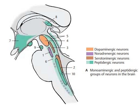
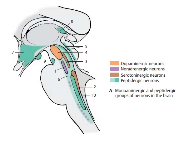
Noradrenergic, dopaminergic, and
sero-toninergic neurons are located in the brainstem.Noradrenergic neuronsform the locus caeruleus (A1) and cell groups in the lateral part
of the reticular formation of the
medulla oblongataand the pons; their fibers project to the hy-pothalamus, to
the limbic system, diffusely into the neocortex, and to the anterior horn and
lateral horn of the spinal cord. Sero-toninergic
neurons lie in theraphe nuclei (A2) , especially in the posteriorraphe nucleus (A3); their fibers project tothe
hypothalamus, to the olfactory epithelium, and to the limbic system.

Dopaminergic neurons make up the com-pact part of the substantia nigra (A4) from where the nigrostri-atal
fibers extend to the striatum.
Peptidergic neurons are found in phylo-genetically older brain regions, namely,
in the central gray of the midbrain (A5), in the reticular formation (A6),
in the hypo-thalamus (A7), in the olfactory bulb, in the
habenular nucleus (A8), interpeduncular nu-cleus (A9), and solitary nucleus (A10).Numerous
peptidergic neurons have also been demonstrated in the cerebral cortex, in the thalamus,
in the striatum, and in the cerebellum. The significance of the
differentpeptides is still largely unclear. It is assumed that they act as cotransmitters and have a modulating
function. Many of these pep-tides are found in other organs as well, suchas the
digestive system (e.g., vasoactive intestinal polypeptide, somatostatin,
cholecystokinin).
Glutamate is often the transmitter
of pro-jection neurons with long axons. Gluta-matergic
neurons are, for example, the
pro-jection neurons of the cerebral cortex, the pyra-midal cells .
GABAergic inhibitory neurons are oftenclassified according to the target structures on
which they form inhibitory synapses. GABAergic basket cells, which form syn-apses with cell bodies, are
distinguished from axo-axonal cells.
The latter develop in-hibitory synapses at the beginning of the axon (initial
segment) of a projection neu-ron. GABAergic neurons often form local cir-cuits
(interneurons). They often contain
pep-tides (see above) and calcium-binding pro-teins apart from GABA as the
classic trans-mitter.

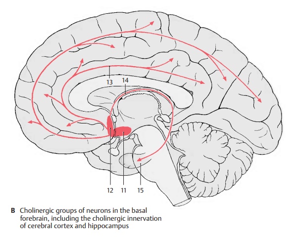
Cholinergic neurons are found in thebrainstem
and also in the basal forebrain.
As in thecase of catecholaminergic neurons, far-reaching projections originate
from circum-scribed groups of cells, for example, in the basal nucleus (B11) and
in certain septal nu-clei (B12) that supply, via fibers in the cingu-late gyrus (limbic gyrus) (B13) and in the fornix (B14),
respectively, large regions ofthe neocortex
and the hippocampus (B15). These ascending cholinergic
projections from the basal forebrain are thought to be associated with processes of learning andmemory. They
are affected in Alzheimer’s dis-ease which
is accompanied by disturbedlearning and memory.
Synthesis, degradation, and
storage of transmitter substances can be influenced by pharmaceuticals. An excess or deficiency intransmitters can be
created in the nerve cells, leading to changes in motor or mental activity.
Changing transmitter synthesis and degradation is not the only way
neurophar-maceuticals can influence the synaptic transmission; they can also
act on the re-ceptors as transmitter agonists or antago-nists.
Related Topics