Chapter: Human Nervous System and Sensory Organs : Basic Elements of the Nervous System
Neuroglia

Neuroglia
The neuroglia (glia, glue) is
the supportingand covering tissue of the
CNS and has all thefunctions of connective tissue: support, metabolite
exchange, and, in pathological processes, digestion of degenerating cells
(phagocytosis) and scar formation. It is of ectodermal
origin. After Nissl staining, onlycell nuclei and cytoplasm are visible;
visual-ization of cellular processes is achieved only by special impregnation
methods and by immunocytochemistry. We distinguish three different types of
neuroglia: astroglia (macroglia), oligodendroglia, and microglia
(A).
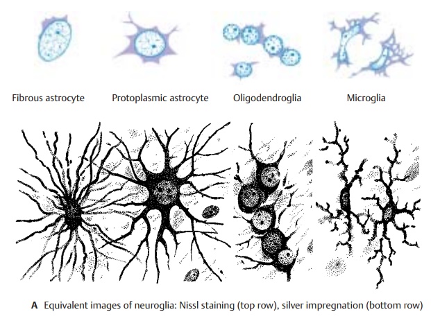
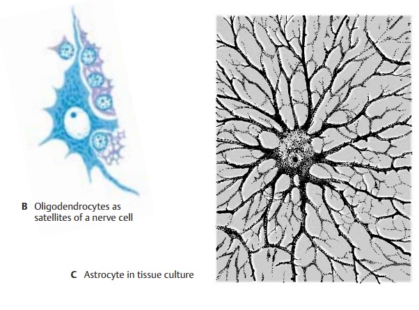
Astrocytes have a large, clear cell nucleusand numerous processes which
give the cell a starlike appearance (A,
C). There are proto-plasmic astrocytes with few processes (usu-ally present in
gray matter) and fibrous astro-cytes with
numerous long processes (pre-dominantly present in white matter). The latter produce
fibers and contain glial fila-ments in cell body and processes. They form glial
scars after damage to the brain tissue. Astrocytes are regarded as supporting
el-ements, since they form a three-dimen-sional scaffold. On the outer surface
of the brain, the scaffold thickens to form a dense fiber felt, the glial limiting membrane, which forms
the outer limit of the ectodermal tissue against the mesenchymal meninges.
Astrocytes extend processes to blood ves-sels and play a role in metabolite
exchange.

In addition, astrocytes play a
decisive part in maintaining the interior
environment of theCNS, particularly the ion
balance. Potassium ions released upon excitation of groups of neurons are
removed from the extracellular space via the network of astrocyte processes.
Astrocytes probably also take up CO2 released by nerve cells and thus keep the
interstitial pH at a constant value of 7.3. Astrocyte processes enclose the
synapses and seal off the synaptic cleft. They also take up neurotransmitters
(uptake and release of GABA via astrocytes have been demon-strated).
Oligodendrocytes have a smaller, darkercell nucleus and only a few,
sparsely branched processes. In the gray matter, they accompany neurons (satellite cells) (B). In the white matter, they lie in rows between the nerve fibers (intrafascicular glia). They pro-duce
and maintain the myelin sheath. In
the peripheral nervous system the myelin sheath is formed by Schwann cells.

Microglial cells have an oval or rodlike cellnucleus and short, branched processes. They exhibit ameboid mobility and can mi-grate within the
brain tissue. In response to tissue destruction, they phagocytose mate-rial (scavenger cells) and round up into spheres (gitter cell).
The frequently expressed view that
micro-glia were not derived from the ectoderm but from the mesoderm (mesoglia) is not sup-ported by evidence.
Related Topics