Chapter: Human Nervous System and Sensory Organs : Basic Elements of the Nervous System
Structure of the Myelin Sheath in the CNS - The Nerve Fiber

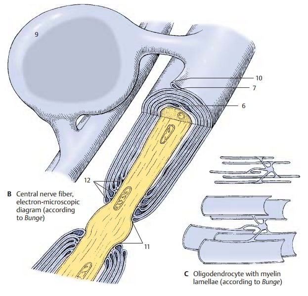
Structure of the Myelin Sheath in the CNS
The myelin sheath in the CNS (B) shows distinct differences as
compared to the myelin sheath of the peripheral nerves . Whereas
the Schwann cell in the PNS myeli-nates only one axon, an oligodendrocyte (B9) in
the CNS myelinates several axons and will later remain connected with several
inter-nodes via cytoplasmic bridges. The extent and shape of the cell becomes
clear when visualizing the internodes as being unfurled (C). The mechanism of the myelination process is unknown. The
external mesaxon forms an external bulge
(B10) starting from the cytoplasmic
bridge. The myelin lamellae terminate at the paranodal region (B11)
(node of Ranvier). As seen in the longitudi-nal section, the innermost lamella
termi-nates first and the outermost lamella covers the remaining endings,
terminating directly at the node of Ranvier. At the ends of the lamellae, the
dense major period lines widen into pockets
filled with cytoplasm (B12). The
axon of the central nerve fiber is completely exposed in the area of the node
of Ranvier. There are no Schmidt–Lanter-man incisures in the CNS.
Related Topics