Chapter: Biotechnology Applying the Genetic Revolution: Immune Technology
The ELISA as a Diagnostic Tool
THE
ELISA AS A DIAGNOSTIC TOOL
The ELISA is used in many
different fields. Diagnostic kits that rely on the ELISA are produced for
clinical diagnosis of human disease, dairy and poultry diseases, and even for
plant diseases. The diagnostic kits are so simple that most require no
laboratory equipment and take as little as 5 minutes. ELISA kits can be used to
detect a particular plant disease by crushing a leaf and smearing the leaf
tissue on the antibody. When the disease-specific antigen reacts with the
antibody, the antibody spot turns blue. In clinical applications, ELISA kits
can detect the presence of minute amounts of pathogenic viruses or bacteria,
even before the pathogen has a chance to cause major damage. Clinical ELISA
kits detect various disease markers. In certain diseases, characteristic
proteins mark the start of disease progression long before the patient exhibits
any symptoms. Detecting such markers can help diagnose and treat a problem
before the disease causes serious damage.

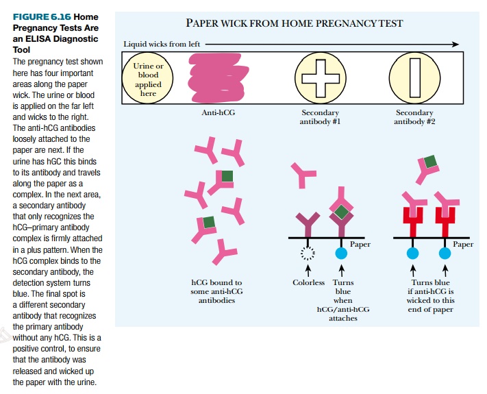
ELISA diagnostic testing is even available for you to try at home. Home pregnancy kits are a simple, over-the-counter ELISA assay for human chorionic gonadotropin (hGC). This is a protein produced by the placenta and secreted into the bloodstream and urine of pregnant women. The actual pregnancy test has four important features (Fig. 6.16). First, the entire test is on a piece of paper that wicks the urine from one end to the other. This paper has three regions: first, a region where anti-hCG antibody is loosely attached to the paper strip; second, a region called the pregnancy window; and finally, a control window. As the urine wicks up the paper strip, any hCG present is bound by the anti-hCG antibody. If the woman is pregnant, the anti-hCG/hCG complex moves up the paper strip. If the woman is not pregnant, the anti-hCG antibody moves up the paper strip alone. (Even if the woman is pregnant, there is excess anti-hCG, and so unbound anti-hCG antibody is always found.) If the woman is pregnant, the anti-hCG/hCG complex reaches the pregnancy window where it binds to secondary antibody 1. This is attached to the paper in the shape of a plus sign and cannot move. The secondary antibody has a color detection system attached to it. When the anti-hCG/hCG complex binds to the secondary antibody it triggers color release and a plus sign forms. The control window contains secondary antibody 2. This recognizes only anti-hCG antibody that is not bound to hCG, so its color is activated whether or not the woman is pregnant.


Related Topics