Chapter: Biotechnology Applying the Genetic Revolution: Immune Technology
Fluorescence Activated Cell Sorting
FLUORESCENCE
ACTIVATED CELL SORTING
As explained in the last
section, fluorescent antibodies are used to find the location of intracellular
proteins. Fluorescent antibodies are also able to bind to surface antigens.
Many cells of the immune system have specific antigens on their surface that
distinguish them from others. Each immune cell can have over 100,000 antigen
molecules on their surface. These surface antigens characterize the different
types of immune cells and are systematically named by assigning them a cluster of differentiation (CD) antigen
number. The antigens were mostly identified before their physiological function
was known. For example, CD4 antigens are associated with T-helper cells, and
CD8 with killer T cells. Monoclonal antibodies are available to label many CD
antigens, especially the most common.
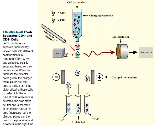
Fluorescence activated cell sorting (FACS) involves the mechanical
separation of a mixture of cells
into different tubes based on their surface antigens (Fig. 6.18). Because the
antibody attaches to the outside of the cell, the cell does not have to be
prepared like above. In this example, helper T cells and killer T cells can be
separated from other white blood cells based on the presence of CD4 or CD8
surface antigens. First, the cell suspension is labeled with monoclonal
antibodies

to the surface antigens of interest. In this example, antibodies to CD4 and CD8 are used. Both antibodies have fluorescent labels that are different for the two antibodies. The labeled cell suspension is loaded into a charging electrode. Drops of liquid containing only one cell are released to the bottom, and the fluorescence detector notes whether or not the drop of liquid is labeled for CD4 or CD8 by the color of its fluorescence. If the drop has an antigen, an electrical charger pulls or pushes the droplet to the right or left, separating the two antigens into separate tubes. If the drop has no antigen in it, it gets no electrical charge and goes into a third tube. Usually two different antibodies are used, but some of the newer FACS machines can sort up to 12 different fluorescently labeled antibodies and can sort up to 300,000 cells per minute.
Flow cytometry is a related technique to analyze
fluorescently labeled cells. As with FACS, cells are labeled with monoclonal
antibodies to cell-surface antigens. The antibodies are conjugated to a variety
of different fluorescent labels, and each antibody is detected based on its
fluorescence.
The cells are loaded into a
charging electrode and released in small droplets. During flow cytometry, the
cells are not sorted and saved; instead the sample of cells is measured and
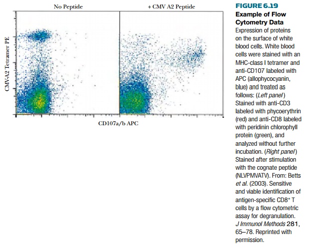
discarded. As the cells pass the detector, the computer records the
fluorescence and plots the number of cells with each of the fluorescent labels.
These are plotted with a small dot representing each of the cells (Fig. 6.19).

Related Topics