Ear - Phonoreceptor | 11th Zoology : Chapter 10 : Neural Control and Coordination
Chapter: 11th Zoology : Chapter 10 : Neural Control and Coordination
Phonoreceptor
Phonoreceptor
The ear is the site of reception of two senses
namely hearing and equilibrium. Anatomically, the ear is divided into three
regions: the external ear, the middle ear and internal ear.
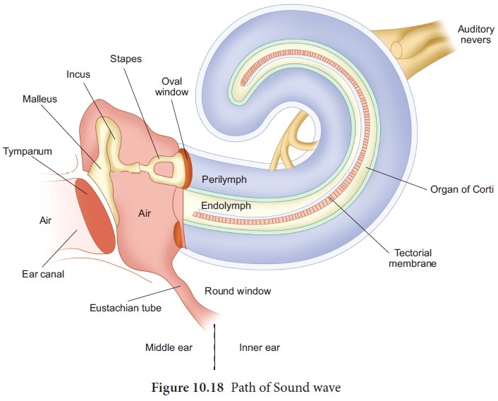
The external ear consists of pinna, external auditory meatus and ear drum. The pinna is flap of elastic cartilage covered by skin.
It collects the sound waves. The external auditory meatus is a curved tube that
extends up to the tympanic membrane [the ear drum]. The tympanic membrane is
composed of connective tissues covered with skin outside and with mucus
membrane inside.
There are very fine hairs and wax producing
sebaceous glands called ceruminous glands in the external auditory meatus.
The combination of hair and the ear wax [cerumen]
helps in preventing dust and foreign particles from entering the ear.
The middle ear is a small air-filled cavity in the temporal bone. It is separated from the external ear by the eardrum and from the internal ear by a thin bony partition; the bony partition contains two small membrane covered openings called the oval window and the round window.
The middle ear contains three ossicles: malleus [hammer bone], incus [anvil bone] and stapes [stirrup bone] which are
attached to one another. The malleus is attached to the tympanic membrane and
its head articulates with the incus which is the intermediate bone lying
between the malleus and stapes. The stapes is attached to the oval window in
the inner ear. The ear ossicles transmit sound waves to the inner ear. A tube
called Eustachian tube connects the middle ear cavity with the pharynx. This
tube helps in equalizing the pressure of air on either sides of the ear drum.
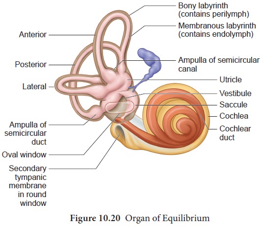
Inner ear is the
fluid filled cavity consisting of two parts, the bony labyrinth and the
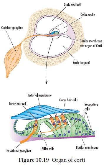
membranous labyrinths. The bony labyrinth consists of three areas: cochlea, vestibule and semicircular canals. The cochlea is a coiled portion consisting
of 3 chambers namely: scala vestibuli
and scala tympani- these two are
filled with perilymph; and the scala media is filled with endolymph. At the base of the cochlea,
the scala vestibule ends at the ‘oval window’ whereas the scala tympani ends at
the ‘round window’ of the middle ear. The chambers scala vestibuli and scala
media are separated by a membrane called Reisner’s membrane whereas the scala
media and scala tympani are separated by a membrane called Basilar membrane (Figure 10.19).
Organ of corti
The organ of
corti (figure.10.19) is a sensory ridge located on the top of the Basilar membrane and it contains numerous hair cells that are arranged in
four rows along the length of the
basilar membrane. Protruding from the apical part of each hair cell is hair
like structures known as stereocilia.
During the conduction of sound wave, stereocilia makes a contact with the stiff
gel membrane called tectorial membrane,
a roof like structure overhanging the organ of corti throughout its length.
Mechanism of hearing
Sound waves entering the external auditory meatus fall on the tympanic membrane. This causes the ear drum to vibrate, and these vibrations are transmitted to the oval window through the three auditory ossicles. Since the tympanic membrane is 17-20 times larger than the oval window, the pressure exerted on the oval window is about 20 times more than that on the tympanic membrane.
This increased pressure
generates pressure waves in the fluid of perilymph. This pressure causes the round
window to alternately bulge outward and inward meanwhile the basilar membrane
along with the organ of Corti move up and down. These movements of the hair
alternately open and close the mechanically gated ion channels in the base of
hair cells and the action potential is propagated to the brain as sound
sensation through cochlear nerve.
Defects of Ear
Deafness may be temporary or permanent. It can be
further classified into conductive deafness and sensory-neural deafness. Possible causes for conductive deafness
may be due to
i.
the blockage of ear canal with earwax,
iii.
Middle ear infection with fluid accumulation
iii.
Restriction of ossicular movement.
In sensory
-neural deafness, the defect may be in the organ of Corti or the auditory
nerve or in the ascending auditory pathways or auditory cortex.
Organ of Equilibrium
Balance is part of a sense called proprioception, which is the ability to sense the position, orientation and movement of the body.
The organ of balance is known as the vestibular
system which is located in the inner ear next to the cochlea. The
vestibular system is composed of a series of fluid filled sacs and
tubules.These sacs and tubules contain endolymph and are kept in the
surrounding perilymph (Figure-10.20). These two fluids, perilymph and
endolymph, respond to the mechanical forces, during changes occurring in body
position and acceleration (Figure 10.21).
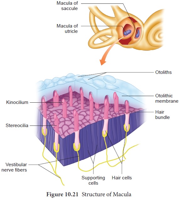
The utricle and saccule are two membranous sacs,
found nearest the cochlea and contain equilibrium receptor regions called maculae that are involved in detecting
the linear movement of the head. The maculae contain the hair cells that act as
mechanorecptors. These hair cells are embeded in a gelatinous otolithic
membrane that contains small calcareous particles called otoliths. This membrane adds weight to the top of the hair cells
and increase the inertia.
The canals that lie posterior and lateral to the
vestibule are semicircular canals; they are anterior, posterior and lateral canals oriented at right angles to
each other. At one end of each
semicircular canal, at its lower end has a swollen area called ampulla.
Each ampulla has a sensory area known as crista ampullaris which is formed of
sensory hair cells and supporting cells. The function of these canals is to
detect rotational movement of the head.
Related Topics