Skin - Olfactory receptors | 11th Zoology : Chapter 10 : Neural Control and Coordination
Chapter: 11th Zoology : Chapter 10 : Neural Control and Coordination
Olfactory receptors
Olfactory receptors
The receptors for taste and smell are the
chemoreceptors. The smell receptors are excited by air borne chemicals that
dissolve in fluids. The yellow coloured patches of olfactory epithelium form
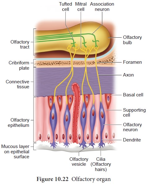
the olfactory organs (figure.10.22) that are located on the roof of the nasal
cavity. The olfactory epithelium is covered by a thin coat of mucus layer below
and olfactory glands bounded connective tissues, above. It contains three types
of cells: supporting cells, Basal cells
and millions of pin shaped olfactory
receptor cells (which are unusual
bipolar cells). The olfactory glands and the supporting cells secrete the mucus. The unmyelinated axons of the
olfactory receptor cells are gathered to form the filaments of olfactory nerve
[cranial nerve I] which synapse with cells of olfactory bulb. The impulse,
through the olfactory nerves, is transmitted to the frontal lobe of the brain
for identification of smell and the limbic system for the emotional responses
to odour.

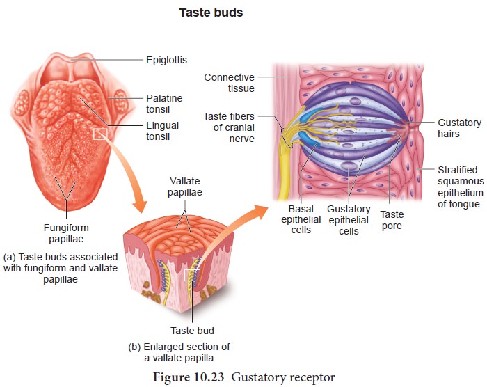
Gustatory receptor: The sense of taste is considered to be the most pleasurable of all senses. The tongue is provided with many small projections called papillae which give the tongue an abrasive feel. Taste buds are located mainly on the papillae which are scattered over the entire tongue surface.
Most taste buds are seen on the tongue (Figure
10.23) few are scattered on the soft palate, inner surface of the cheeks,
pharynx and epiglottis of the larynx. Taste buds are flask-shaped and consist
of 50 – 100 epithelial cells of two major types.

Gustatory
epithelial cells (taste cells) and Basal epithelial cells (Repairing cells) Long microvilli called gustatory hairs project from the tip of
the gustatory cells and extends through a taste pore to the surface of the
epithelium where they are bathed by saliva. Gustatory- hairs are the sensitive
portion of the gustatory cells and they have sensory dendrites which send the
signal to the brain. The basal cells that act as stem cells, divide- and
differentiate into new gustatory cells (Figure 10.23).
Skin-Sense of touch
Skin is the sensory organ of touch and is also the largest sense organ. This sensation comes from millions of microscopic sensory receptors located all over the skin and associated with the general sensations of contact, pressure, heat, cold and pain. Some parts of the body, such as the finger tips have a large number of these receptors, making them more sensitive. Some of the sensory receptors present in the skin (Figure 10.24) are:

•
Tactile
merkel disc is light touch receptor lying in the deeper layer
of epidermis.
•
Hair
follicle receptors are light touch receptors lying around the hair follicles.
•
Meissner’s
corpuscles are small light pressure receptors found just
beneath the epidermis in the dermal
papillae. They are numerous in hairless skin areas such as finger tips and
soles of the feet.
•
Pacinian
corpuscles are the large egg shaped receptors found scattered deep in the dermis and monitoring
vibration due to pressure. It allows to detect different textures, temperature,
hardness and pain
•
Ruffini
endings which lie in the dermis responds-
to continuous pressure.
• Krause end bulbs are thermoreceptors that sense temperature.
Melanocytes are the cells responsible for producing
the skin pigment, melanin, which gives skin its colour and protects it from the
sun's UV rays. Vitiligo (Leucoderma) is a condition in which the melanin
pigment is lost from areas of the skin, causing white patches, often with no
clear cause. Vitiligo is not contagious. It can affect people of any age,
gender, or ethnic group. The patches appear when melanocytes fails to synthesis
melanin pigment.
Related Topics