Chapter: 11th Zoology : Chapter 10 : Neural Control and Coordination
Central neural system (CNS)
Central neural system (CNS)
The CNS includes the brain and the spinal cord,
which are protected by the bones of the skull and vertebral column. During its
embryonic development, CNS develops from the ectoderm.
1. Brain
The brain acts as the command and control system.
It is the site of information processing. It is located in the cranial cavity
and is covered by three cranial meninges. The outer thick layer is Duramater
which lines the inner surface of the cranial cavity; the median thin layer is
Arachnoid mater which is separated from the duramater by a narrow subdural
space. The innermost layer is Piamater which is closely adhered to the brain
but separated from the arachnoid mater by the subarachnoid space. The brain is
divided into three major regions: Forebrain, Midbrain and Hindbrain.
Fore Brain
It comprises the following regions: Cerebrum and
Diencephalon. Cerebrum is the ‘seat of intelligence’ and forms the major part
of the brain. The cerebrum consists of an outer cortex, inner medulla and basal
nuclei. The superficial region of the cerebrum is called cerebral cortex, which
looks grey due to the presence of unmyelinated nerve cells. Cerebral cortex
consists of neuronal cell body, dendrites, associated glial and blood vessels.
The surface of the cerebrum shows many convolutions (folds) and grooves. The
folds are called gyri (singular gyrus); the shallow grooves between the gyri
are called sulci (singular sulcus) and deep grooves are called fissures. These
sulci and gyri increase the surface area of the cerebral cortex. Several sulci
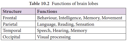
divide the cerebrum into eight lobes: a pair of frontals, parietals, temporals and occipital lobes (Figure10.7 & Table 10.2).

A median longitudinal fissure divides the cerebrum
longitudinally into two cerebral hemispheres (Figure 10.7). A transverse
fissure separates the cerebral hemispheres from the cerebellum. The
Cerebral
cortex has three functional areas namely sensory
areas occur in the parietal, temporal and occipital lobes of the cortex.
They receive and interpret the sensory impulses. Motor area of the cortex which controls voluntary muscular
movements lies in the posterior part
of the frontal lobes. The areas other than sensory and motor areas are called Association areas that deal with
integrative functions such as memory, communications, learning and reasoning.
Inner to the cortex is medulla which
is white in colour and actsas a nerve tract between the cortex and the
diencephalon.
Diencephalon
consists
largely of following three paired structures.
Epithalamus forms
the roof of the diencephalon and it is a non-nervous tissue. The anterior part of epithalamus is
vascular and folded to form the anterior choroid
plexus. Just behind the choroid
plexus, the epithalamus forms a short stalk which ends in a rounded body called pineal
body which secretes the hormone, melatonin
which regulates sleep and wake cycle.
Thalamus is composed of grey mater which serves as a relay centre for impulses between the spinal cord, brain stem and
cerebrum. Within the thalamus, information is sorted and edited and plays a key
role inlearning and memory. It is a major coordinating centre for sensory and
motor signalling.
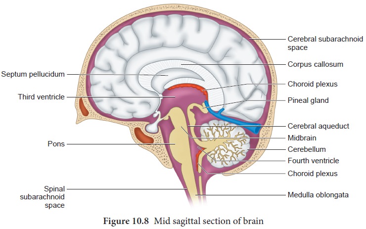
Hypothalamus
forms the
floor of the diencephalon. The downward extension of the hypothalamus, the infundibulum
connect sthe hypothalamus with the pituitary gland. The hypothalamus contains a pair
of small rounded body called mammillary
bodies that are involved in olfactory reflexes and emotional responses to
odour. Hypothalamus maintains homeostasis and has many centres which control
the body temperature, urge for eating and drinking. It also contains a group of
neurosecretory cells which secrete the hypothalamic hormones. Hypothalamus also
acts as the satiety centre.
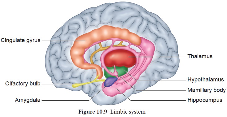
Limbic system
The inner part of the cerebral hemisphere
constitutes the limbic system. The main components of limbic system are olfactory bulbs, cingulate gyrus,
mammillary body, amygdala,
hippocampus and hypothalamus. The limbic system is called ‘emotional brain’ because it plays a
primary role in the regulation of pleasure, pain, anger, fear, sexual feeling
and affection. The hippocampus and amygdala also play a role in memory (Figure
10.9).
Brain
stem is the part of the brain between the spinal cord and the diencephalon.
It consists of mid brain, pons
varolii and medulla oblongata (Figure 10.10).
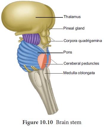
Mid brain
The mid brain is located between the diencephalon
and the pons. The lower portion of
the midbrain consists of a pair of longitudinal bands of nervous tissue called
cerebral peduncles which relay impulses back and forth between cerebrum,
cerebellum, pons and medulla. The dorsal portion of the midbrain consists of
four rounded bodies called corpora
quadrigemina which acts as a reflex centre for vision and hearing.
Hind brain
Rhombencephalon forms the hind brain. It comprises
of cerebellum, pons varolii and medulla oblongata. Cerebellum is the second
largest part of the brain. It consists of two cerebellar hemispheres and
central worm shaped part, the vermis. The cerebellum controls and coordinates
muscular movements and body equilibrium. Any damage to cerebellum often results
in uncoordinated voluntary muscle movements.
Pons
varoli lies infront of the cerebellum between the midbrain and the medulla oblongata. The nerve fibres in the
pons varolii form a bridge between the two cerebellar hemispheres and connect
the medulla oblongata with the other region of the brain. The respiratory
nuclei found in the pons cooperate with the medulla to control respiration.
Medulla
oblongata forms the posterior most part of the brain. It connects the spinal cord with various parts of the brain.
It receives and integrates signals from spinal cord and sends it to the
cerebellum and thalamus. Medulla contains vital centres that control cardio
vascular reflexes, respiration and gastric secretions.
Ventricles of the brain
The brain has four hollow, fluid filled spaces. The
C- shaped space found inside each cerebral hemisphere forms the lateral
ventricles I and II which are separated from each other by a thin membrane
called the septumpellucidum. Each lateral ventricle communicates with the
narrow III ventricle in the diencephalon through an opening called
interventricular foramen (foramen of Monro). The ventricle III is continuous
with the ventricle IV in the hind brain through a canal called aqueduct of
Sylvius (cerebral aqueduct). Choroid plexus is a network of blood capillaries
found in the roof of the ventricles and forms cerebro spinal fluid (CSF) from
the blood. CSF provides buoyancy to the CNS structures; CSF acts as a shock
absorber for the brain and spinal cord; it nourishes the brain cells by
transporting constant supply of food and oxygen; it carries harmful metabolic
wastes from the brain to the blood; and maintains a constant pressure inside
the cranial vessels.
2. Spinal cord
The spinal cord is a long, slender, cylindrical
nervous tissue. It is protected by the vertebral column and surrounded by the
three membranes as in the brain. The spinal cord that extends from the brain
stem into the vertebral canal of the vertebral column up to the level of 1st or
2nd lumbar vertebra. So the nerve roots of the remaining nerves are greatly
elongated to exit the vertebral column at their appropriate space. The thick
bundle of elongated nerve roots within the lower vertebral canal is called the cauda equina (horse’s tail) because of its appearance.
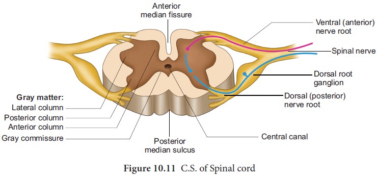
In the cross section of spinal cord (Figure 10.11),
there are two indentations: the posterior median sulcus and the anterior median
fissure. Although there might be slight variations, the cross section of spinal
cord is generally the same throughout its length. In contrast to the brain, the
grey matter in the spinal cord forms an inner butterfly shaped region
surrounded by the outer white matter.
The grey matter consists of neuronal cell bodies and their dendrites,
interneurons and glial cells. White matter consists of bundles of nerve fibres.
In the center of the grey matter there is a central canal
which is filled with CSF. Each half of the grey matter is divided into a dorsal horn, a ventral horn and a
lateral horn.
The dorsal horn contains cell bodies of
interneurons on which afferent neurons terminate. The ventral horn contains
cell bodies of the efferent motor neurons supplying the skeletal muscle.
Autonomic nerve fibres, supplying cardiac and smooth muscles and exocrine
glands, originate from the cell bodies found in the lateral horn. In the white
matter, the bundles of nerve fibres form two types of tracts namely ascending tracts which carry sensory
impulses to the brain and descending
tracts which carry motor impulses from the brain to the spinal nerves at
various levels of the spinal cord. The spinal cord shows two enlargements, one
in the cervical region and another one in the lumbosacral region. The cervical
Related Topics