Chapter: Human Nervous System and Sensory Organs : Spinal Cord and Spinal Nerves
Overview of Spinal Cord and Spinal Nerves

Spinal Cord and Spinal Nerves
Overview
The spinal cord, or spinal medulla, is located in the channel of the spinal column,
the vertebral canal, and is
surrounded by the cerebrospinal fluid.
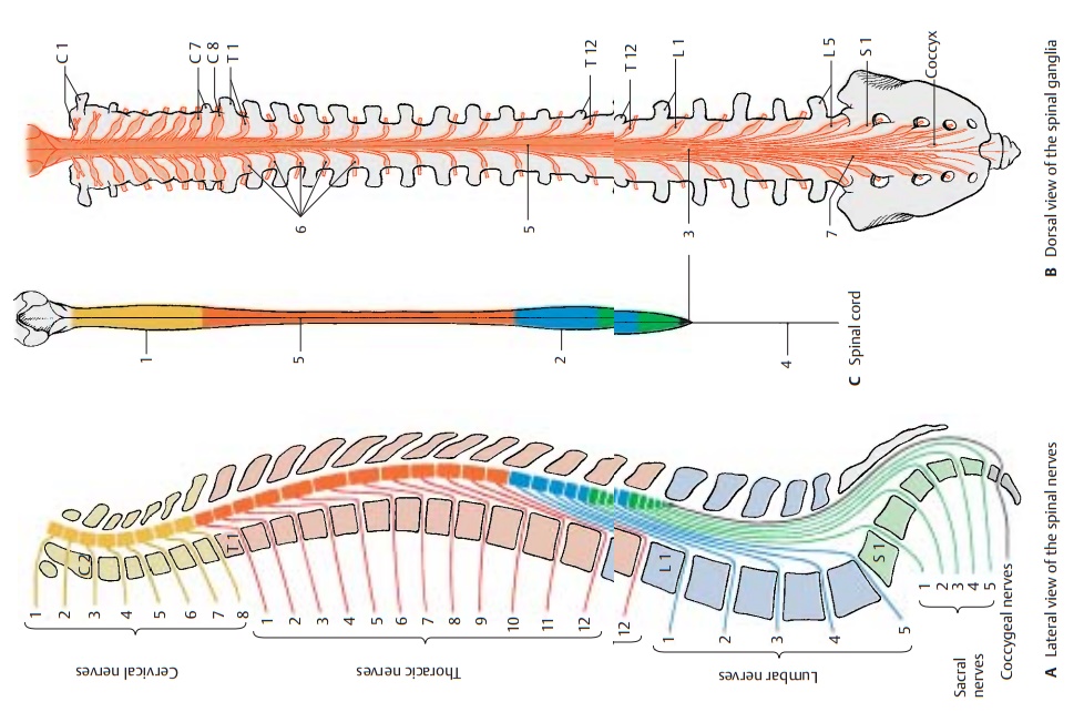
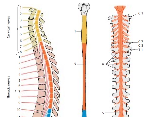
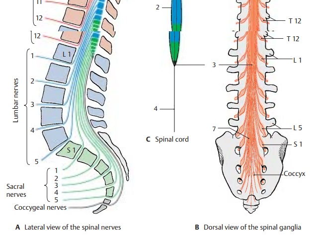
It has two spindle-shaped swellings: one in the neck region, the cervical enlargement (C1), and one in the lumbar region, the lumbar enlargement (C2). At the lower end, the spinal cord
tapers into the medullary cone (BC3) and ends as a thin thread, the terminal filament (C4). The ante-rior median
fissure at the ventral side and the
posterior median sulcus (BC5) at
the dorsalside mark the boundaries between the two symmetrical halves of the
spinal cord. Nerve fibers enter dorsolaterally and emerge ven-trolaterally at
both sides of the spinal cord and unite to form the dorsal roots, posteriorroots,
and theventral roots, anterior roots. Theroots join to form
short nerve trunks of 1 cm in length, the spinal
nerves. Intercalated into the posterior roots are spinal ganglia (B6)
containing sensory nerve cells. Only
the posterior roots of the first cervical spinal nerves do not have a spinal
ganglion, or only a rudimentary one.
In humans, there are 31 pairs of spinal nerves which emerge
through the intervertebralforamina from
the vertebral canal. Each spi-nal
nerve pair supplies one body segment. The spinal cord itself is unsegmented.
The impression of segmentation is created by the bundling of nerve fibers
emerging from the foramina.
The
spinal nerves are subdivided into cervi-cal
nerves, thoracic nerves, lumbar nerves, sacral nerves, and
coccygeal nerves (A). There are
8 pairs of cervical nerves (C1 –
C8) (the first pair emerges between occipital bone and atlas)
12airs of thoracic nerves (T1 –
T12) (thefirst pair emerges between the first and second thoracic vertebrae
5 pairs of lumbar nerves (L1 –
L5) (the firstpair emerges between the first and sec-ond lumbar vertebrae)
5 pairs of sacral nerves (S1 –
S5) (the firstpair emerges through the upper sacral foramina)
one pair of coccygeal nerves (emerging be-tween the first and
second coccygeal vertebrae)


Spinal
cord and vertebral canal are initially of the same length so that each spinal
nerve emerges from the foramen lying at its own level. During development,
however, the vertebral column increases much more in length than does the
spinal cord. As a result, the lower end of the spinal cord moves further up in
relation to the surrounding vertebrae. In the newborn, the lower end of the
spinal cord lies at the level of the third lumbar vertebra, and in the adult,
at the level of the first lumbar or twelfth thoracic vertebra. Thus, the spinal
nerves no longer emerge at their levels of origin; instead, their roots run
down a certain distance within the vertebral canal to their foramen where they
emerge. The more caudally the roots originate from the spinal cord, the longer
their run within the vertebral canal. The levels where the spinal nerves emerge
are therefore no longer identical with the corresponding levels of the spinal
cord.
From the
medullary cone (BC3) onward, the
vertebral canal contains only a dense mass of descending spinal roots, known as
the cauda equina (tail of a horse) (B7).
Related Topics